![All lanes : Anti-EIF3S4 antibody [EPR16146] - N-terminal (ab191422) at 1/10000 dilutionLane 1 : 293T cell lysateLane 2 : Molt-4 cell lysateLane 3 : HepG2 cell lysateLane 4 : K562 cell lysateLysates/proteins at 20 µg per lane.SecondaryGoat Anti-Rabbit IgG, (H+L), Peroxidase conjugated at 1/1000 dilution](http://www.bioprodhub.com/system/product_images/ab_products/2/sub_2/12783_ab191422-230789-ab1914221.jpg)
All lanes : Anti-EIF3S4 antibody [EPR16146] - N-terminal (ab191422) at 1/10000 dilutionLane 1 : 293T cell lysateLane 2 : Molt-4 cell lysateLane 3 : HepG2 cell lysateLane 4 : K562 cell lysateLysates/proteins at 20 µg per lane.SecondaryGoat Anti-Rabbit IgG, (H+L), Peroxidase conjugated at 1/1000 dilution
![All lanes : Anti-EIF3S4 antibody [EPR16146] - N-terminal (ab191422) at 1/1000 dilutionLane 1 : PC-12 cell lysateLane 2 : NIH/3T3 cell lysateLysates/proteins at 10 µg per lane.SecondaryGoat Anti-Rabbit IgG, (H+L), Peroxidase conjugated at 1/1000 dilution](http://www.bioprodhub.com/system/product_images/ab_products/2/sub_2/12784_ab191422-230788-ab1914222.jpg)
All lanes : Anti-EIF3S4 antibody [EPR16146] - N-terminal (ab191422) at 1/1000 dilutionLane 1 : PC-12 cell lysateLane 2 : NIH/3T3 cell lysateLysates/proteins at 10 µg per lane.SecondaryGoat Anti-Rabbit IgG, (H+L), Peroxidase conjugated at 1/1000 dilution

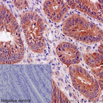
Immunohistochemical analysis of paraffin-embedded human colonic carcinoma tissue sections labeling EIF3S4 using ab191422 at a 1/50 dilution. A ready to use HRP polymer for Rabbit IgG was used as the secondary. Hematoxylin counterstain. Negative control uses PBS instead of primary antibody.

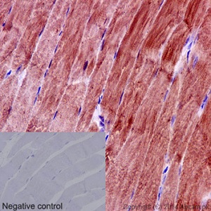
Immunohistochemical analysis of paraffin embedded rat skeletal muscle tissue sections labeling EIF3S4 using ab191422 at a 1/50 dilution. A ready to use HRP polymer for Rabbit IgG was used as the secondary. Hematoxylin counterstain. Negative control uses PBS instead of primary antibody.

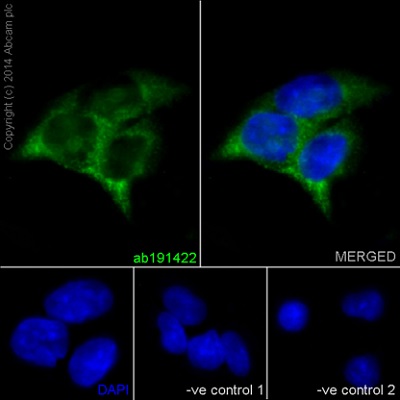
Immunofluorescent analysis of 4% paraformaldehyde fixed HepG2 cells labeling EIF3S4 using ab191422 at a 1/150 dilution. A Goat anti rabbit IgG (Alexa Fluor®488) (ab150077) was used as the secondary at a 1/200 dilution. Counterstain DAPI. Cells were permeabilized using 0.1% Triton X-100. The two negative controls: Primary ab concentration (anti-EIF3G), Secondary ab (Goat anti mouse IgG (Alexa Fluor®594)) is 1/400 dilution.

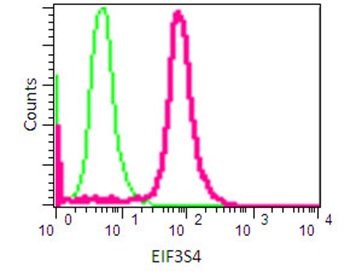
Flow Cytometry analysis of 2% paraformaldehyde fixed 293T cells labeling EIF3S4 using ab191422 at a 1/80 dilution (pink). Goat anti rabbit IgG (FITC) used as the secondary at a 1/150 dilution. Isotype control Rabbit monoclonal IgG (green).
![All lanes : Anti-EIF3S4 antibody [EPR16146] - N-terminal (ab191422) at 1/10000 dilutionLane 1 : 293T cell lysateLane 2 : Molt-4 cell lysateLane 3 : HepG2 cell lysateLane 4 : K562 cell lysateLysates/proteins at 20 µg per lane.SecondaryGoat Anti-Rabbit IgG, (H+L), Peroxidase conjugated at 1/1000 dilution](http://www.bioprodhub.com/system/product_images/ab_products/2/sub_2/12783_ab191422-230789-ab1914221.jpg)
![All lanes : Anti-EIF3S4 antibody [EPR16146] - N-terminal (ab191422) at 1/1000 dilutionLane 1 : PC-12 cell lysateLane 2 : NIH/3T3 cell lysateLysates/proteins at 10 µg per lane.SecondaryGoat Anti-Rabbit IgG, (H+L), Peroxidase conjugated at 1/1000 dilution](http://www.bioprodhub.com/system/product_images/ab_products/2/sub_2/12784_ab191422-230788-ab1914222.jpg)


