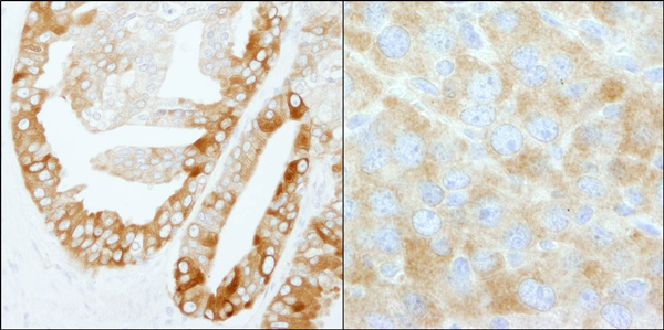
Immunohistochemistry (Formalin/PFA-fixed paraffin-embedded sections) analysis of human prostate carcinoma (left) and mouse renal cell carcinoma (right) tissues labelling Fatty Acid Synthase with ab99359 at 1/200 (1µg/ml). Detection: DAB.
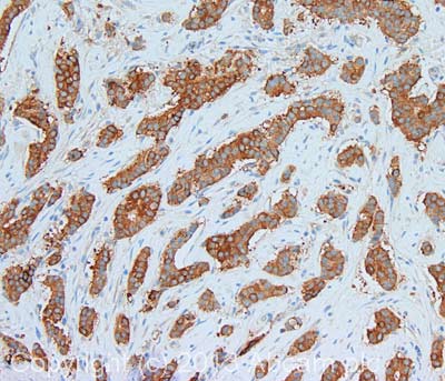
IHC image of Fatty Acid Synthase staining in human breast adenocarcinoma formalin fixed paraffin embedded tissue section, performed on a Leica Bond system using the standard protocol F. The section was pre-treated using heat mediated antigen retrieval with sodium citrate buffer (pH6, epitope retrieval solution 1) for 20 mins. The section was then incubated with ab99359, 1µg/ml, for 15 mins at room temperature and detected using an HRP conjugated compact polymer system. DAB was used as the chromogen. The section was then counterstained with haematoxylin and mounted with DPX.For other IHC staining systems (automated and non-automated) customers should optimize variable parameters such as antigen retrieval conditions, primary antibody concentration and antibody incubation times.
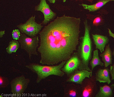
ICC/IF image of ab99359 stained A549 cells. The cells were 4% formaldehyde fixed (10 min) and then incubated in 1%BSA / 10% normal goat serum / 0.3M glycine in 0.1% PBS-Tween for 1h to permeabilise the cells and block non-specific protein-protein interactions. The cells were then incubated with the antibody (ab99359, 1µg/ml) overnight at +4°C. The secondary antibody (green) was ab96899, DyLight® 488 goat anti-rabbit IgG (H+L) used at a 1/250 dilution for 1h. Alexa Fluor® 594 WGA was used to label plasma membranes (red) at a 1/200 dilution for 1h. DAPI was used to stain the cell nuclei (blue) at a concentration of 1.43µM.
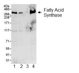
All lanes : Anti-Fatty Acid Synthase antibody (ab99359) at 0.04 µg/mlLane 1 : HeLa whole cell lysate at 50 µgLane 2 : HeLa whole cell lysate at 15 µgLane 3 : HeLa whole cell lysate at 5 µgLane 4 : 293T whole cell lysate at 50 µgdeveloped using the ECL technique
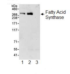
ab99359 at 0.1 µg/ml detecting Fatty Acid Synthase in HeLa whole cell lysate by western blot analysis following immunoprecipitation. Detection utilised ECL with a 30 second exposure. For immunoprecipitation, ab99359 was used at at 3 µg/mg lysate; 1 mg of lysate was used for IP and 20% of IP was loaded. Lane 1; IP using ab99358 (a rabbit anti-Fatty Acid Synthase antibody which recognizes a upstream epitope). Lane 2; IP using ab99359. Lane 3; IP using control IgG.