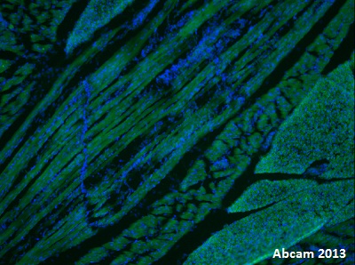
IHC-Fr image of Ferritin, mitochondrial staining on mouse skeletal muscle using ab66111 (1: 100). The sections were fixed in 4% PFA and the section was permeabilized using 0.1% TritonX in 0.1% PBS. The section was then blocked using 10% conkey serum for 1 hour at 24°C. The primary antibody (1:100) was incubated at 4°C for 24 hours and the secondary antibody used was donkey polyclonal to rabbit IgG conjugated to Alexa Fluor 488 (1:1000).

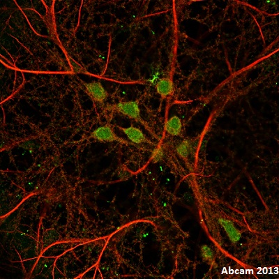
ICC/IF image of Ferritin, mitochondrial staining on mouse hippocampal culture using ab66111 (1: 100). The cells were fixed in 4% PFA and the section was permeabilized using 0.1% TritonX in 0.1% PBS. The cells was then blocked using 10% donkey serum for 1 hour at 24°C. The primary antibody (1:100) was incubated at 4°C for 24 hours and the secondary antibody used was donkey polyclonal to rabbit IgG conjugated to Alexa Fluor 488 (1:1000).

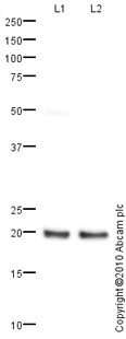
All lanes : Anti-Ferritin, mitochondrial antibody (ab66111) at 1 µg/mlLane 1 : Liver (Human) Tissue Lysate - adult normal tissue (ab29889)Lane 2 : Brain (Human) Tissue Lysate - adult normal tissue (ab29466)Lysates/proteins at 10 µg per lane.SecondaryGoat polyclonal to Rabbit IgG - H&L - Pre-Adsorbed (HRP) at 1/3000 dilutiondeveloped using the ECL techniquePerformed under reducing conditions.


