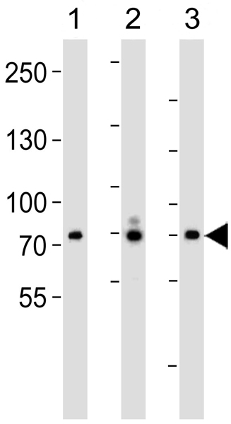
All lanes : Anti-FGFR4 antibody (ab5481) at 1/1000 dilutionLane 1 : SH-SY5Y cell lysateLane 2 : 293 cell lysateLane 3 : Raji cell lysateLysates/proteins at 35 µg per lane.SecondaryHRP-conjugated goat anti-rabbit IgG (H+L) at 1/5000 dilution
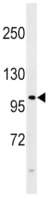
Anti-FGFR4 antibody (ab5481) + 293 cell lysate at 35 µg
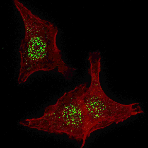
Immunocytochemistry/Immunofluorescence analysis of HeLa cells labelling FGFR4 with ab5481 at 1/200. Cells were fixed with 4% paraformaldehyde for 20 minutes and permeabilized with 0.2% Triton X-100 for 30 minutes. Cells were incubated with the primary antibody for 2 hours at room temperature. An Alexa Fluor® 488 conjugated donkey anti-rabbit IgG (green) was used as the secondary antibody (1/1000, 1 hour). Nuclei were counterstained with Hoechst 33342 (blue) (10 μg/ml, 5 minutes).

Immunohistochemistry (Formalin/PFA-fixed paraffin-embedded sections) analysis of human hepatocarcinoma tissue labelling FGFR4 with ab5481. A peroxidase-conjugated anti-rabbit IgG was used as the secondary antibody, followed by AEC staining.
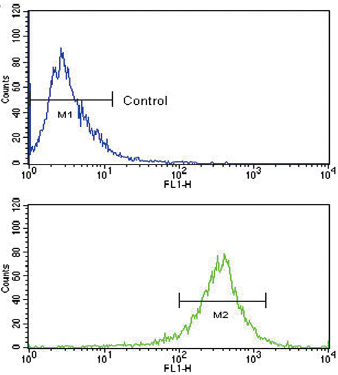
Flow cytometry analysis of WiDr cells labelling FGFR4 (green) with ab5481 compared to a negative control. A FITC-conjugated goat anti-rabbit IgG was used as the secondary antibody.