![Overlay histogram showing SH-SY5Y cells stained with ab130165 (red line). The cells were fixed with 4% paraformaldehyde (10 min)/ and then permeabilized with 0.1% PBS-Tween for 20 min. The cells were then incubated in 1x PBS / 10% normal goat serum / 0.3M glycine to block non-specific protein-protein interactions followed by the antibody (ab130165, 0.1μg/1x106 cells) for 30 min at 22°C. The secondary antibody used was Alexa Fluor® 488 goat anti-mouse IgG (H&L) (ab150113) at 1/2000 dilution for 30 min at 22°C. Isotype control antibody (black line) was mouse IgG1 [ICIGG1] (ab91353, 1μg/1x106 cells) used under the same conditions. Unlabelled sample (blue line) was also used as a control. Acquisition of >5,000 events were collected using a 20mW Argon ion laser (488nm) and 525/30 bandpass filter. This antibody gave a positive signal in SH-SY5Y cells fixed with 80% methanol (5 min)/permeabilized with 0.1% PBS-Tween for 20 min used under the same conditions.](http://www.bioprodhub.com/system/product_images/ab_products/2/sub_2/20027_ab130165-4-ab130165FC.jpg)
Overlay histogram showing SH-SY5Y cells stained with ab130165 (red line). The cells were fixed with 4% paraformaldehyde (10 min)/ and then permeabilized with 0.1% PBS-Tween for 20 min. The cells were then incubated in 1x PBS / 10% normal goat serum / 0.3M glycine to block non-specific protein-protein interactions followed by the antibody (ab130165, 0.1μg/1x106 cells) for 30 min at 22°C. The secondary antibody used was Alexa Fluor® 488 goat anti-mouse IgG (H&L) (ab150113) at 1/2000 dilution for 30 min at 22°C. Isotype control antibody (black line) was mouse IgG1 [ICIGG1] (ab91353, 1μg/1x106 cells) used under the same conditions. Unlabelled sample (blue line) was also used as a control. Acquisition of >5,000 events were collected using a 20mW Argon ion laser (488nm) and 525/30 bandpass filter. This antibody gave a positive signal in SH-SY5Y cells fixed with 80% methanol (5 min)/permeabilized with 0.1% PBS-Tween for 20 min used under the same conditions.
![All lanes : Anti-FMRP antibody [4G9] (ab130165) at 1/500 dilutionLane 1 : Jurkat cell lysatesLane 2 : K562 cell lysates](http://www.bioprodhub.com/system/product_images/ab_products/2/sub_2/20028_FMRP-Primary-antibodies-ab130165-1.jpg)
All lanes : Anti-FMRP antibody [4G9] (ab130165) at 1/500 dilutionLane 1 : Jurkat cell lysatesLane 2 : K562 cell lysates
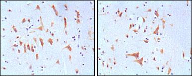
ab130165, at 1/200 dilution, staining FMRP in paraffin-embedded Human brain tissue by Immunohistochemistry, using DAB detection.
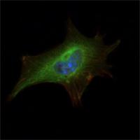
ab130165, at 1/200 dilution, staining FMRP in NIH 3T3 cells by Immunofluorescence (green). DRAQ5 fluorescent DNA dye (blue) and Actin filaments have been labeled with Alexa Fluor-555 phalloidin.