
All lanes : Anti-FOXO1A (phospho S256) antibody (ab26651) at 1 µg/mlLane 1 : Ovary (Human) Tissue LysateLane 2 : Ovary (Human) Tissue Lysate with Human FOXO1A (phospho S256) peptide (ab27886) at 1 µg/mlLane 3 : Ovary (Human) Tissue Lysate with Human FOXO1A peptide (ab27887) at 1 µg/mlLysates/proteins at 20 µg per lane.SecondaryIRDye 680 Conjugated Goat Anti-Rabbit IgG (H+L) at 1/10000 dilutionPerformed under reducing conditions.
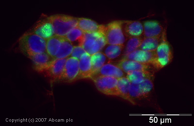
ICC/IF image of ab26651 stained human HEK 293 cells. The cells were PFA fixed (10 min), permabilised in TBS-T (20 min) and incubated with the antibody (ab26651, 1µg/ml) for 1h at room temperature. 1%BSA / 10% normal goat serum / 0.3M glycine was used to quench autofluorescence and block non-specific protein-protein interactions. The secondary antibody (green) was Alexa Fluor® 488 goat anti-rabbit IgG (H+L) used at a 1/1000 dilution for 1h. Alexa Fluor® 594 WGA was used to label plasma membranes (red). DAPI was used to stain the cell nuclei (blue).
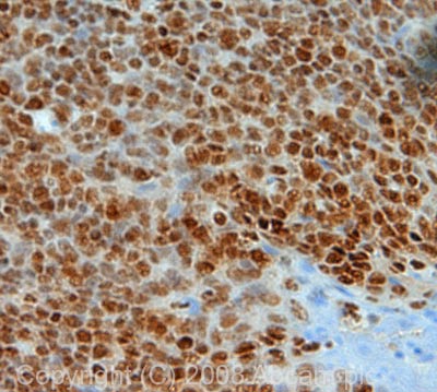
IHC image of FOXO1A (phospho S256) staining in human tonsil FFPE section, performed on a BondTM system using the standard protocol F. The section was pre-treated using heat mediated antigen retrieval with sodium citrate buffer (pH6, epitope retrieval solution 1) for 20 mins. The section was then incubated with ab26651, 1µg/ml, for 8 mins at room temperature and detected using an HRP conjugated compact polymer system. DAB was used as the chromogen. The section was then counterstained with haematoxylin and mounted with DPX.
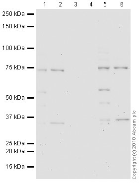
All lanes : Anti-FOXO1A (phospho S256) antibody (ab26651) at 1 µg/mlLane 1 : A431 (Human epithelial carcinoma cell line) Nuclear Lysate - EGF treated (ab14635)Lane 2 : NIH 3T3 (Mouse embryonic fibroblast cell line) Whole Cell LysateLane 3 : A431 (Human epithelial carcinoma cell line) Nuclear Lysate - EGF treated (ab14635) with Human FOXO1A (phospho S256) peptide (ab27886) at 1 µg/mlLane 4 : NIH 3T3 (Mouse embryonic fibroblast cell line) Whole Cell Lysate with Human FOXO1A (phospho S256) peptide (ab27886) at 1 µg/mlLane 5 : A431 (Human epithelial carcinoma cell line) Nuclear Lysate - EGF treated (ab14635) with Human FOXO1A peptide (ab27887) at 1 µg/mlLane 6 : NIH 3T3 (Mouse embryonic fibroblast cell line) Whole Cell Lysate with Human FOXO1A peptide (ab27887) at 1 µg/mlLysates/proteins at 20 µg per lane.SecondaryGoat polyclonal to Rabbit IgG - H&L - Pre-Adsorbed (HRP) at 1/3000 dilutionPerformed under reducing conditions.