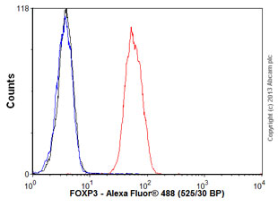
Overlay histogram showing Jurkat cells stained with ab128800 (red line). The cells were fixed with 80% methanol (5 min) and then permeabilized with 0.1% PBS-Tween for 20 min. The cells were then incubated in 1x PBS / 10% normal goat serum / 0.3M glycine to block non-specific protein-protein interactions followed by the antibody (ab128800, 0.1μg/1x106 cells) for 30 min at 22ºC. The secondary antibody used was Alexa Fluor® 488 goat anti-rabbit IgG (H&L) (ab150077) at 1/2000 dilution for 30 min at 22ºC. Isotype control antibody (black line) was rabbit IgG (polyclonal) (0.1μg/1x106 cells) used under the same conditions. Unlabelled sample (blue line) was also used as a control. Acquisition of >5,000 events were collected using a 20mW Argon ion laser (488nm) and 525/30 bandpass filter.
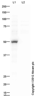
All lanes : Anti-FOXP3 antibody (ab128800) at 1 µg/mlLane 1 : HEK293 Whole Cell Lysate Overexpressing FOXP3 at 0.1 µgLane 2 : HEK293 (Human embryonic kidney cell line) Whole Cell Lysate at 10 µgSecondaryGoat Anti-Rabbit IgG H&L (HRP) (ab97051) at 1/10000 dilutiondeveloped using the ECL techniquePerformed under reducing conditions.
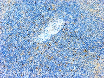
IHC image of FOXP3 staining in Mouse spleen formalin fixed paraffin embedded tissue section, performed on a Leica BondTM system using the standard protocol B. The section was pre-treated using heat mediated antigen retrieval with EDTA based pH 9.0 solution (epitope retrieval solution 2) for 20 mins. The section was then incubated with ab128800, 0.7µg/ml, for 15 mins at room temperature. A Goat anti-Rabbit biotinylated secondary antibody was used to detect the primary, and visualized using an HRP conjugated ABC system. DAB was used as the chromogen. The section was then counterstained with haematoxylin and mounted with DPX. For other IHC staining systems (automated and non-automated) customers should optimize variable parameters such as antigen retrieval conditions, primary antibody concentration and antibody incubation times.