All lanes : Anti-GAB1 antibody (ab59362) at 1/500 dilutionLane 1 : Extracts from NIH/3T3 cells with no immunizing peptideLane 2 : Extracts from NIH/3T3 cells with immunizing peptideObserved band size : 77 kDa (why is the actual band size different from the predicted?)

ab59362, at a 1/50 dilution, staining human GAB1 in breast carcinoma, using Immunohistochemistry, Paraffin embedded tissue, in the absence of the immunizing peptide (left image) and in the presence of the immunizing peptide (right image).

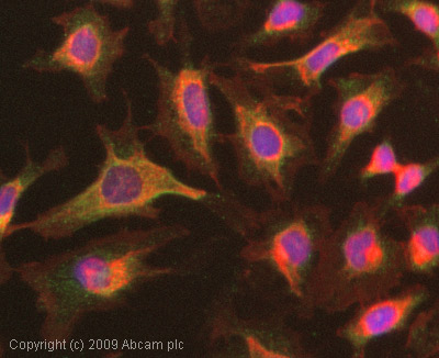
ICC/IF image of ab59362 stained HeLa cells. The cells were 100% methanol fixed (5 min) and then incubated in 1%BSA / 10% normal goat serum / 0.3M glycine in 0.1% PBS-Tween for 1h to permeabilise the cells and block non-specific protein-protein interactions. The cells were then incubated with the antibody (ab59362, 1µg/ml) overnight at +4°C. The secondary antibody (green) was Alexa Fluor® 488 goat anti-rabbit IgG (H+L) used at a 1/1000 dilution for 1h. Alexa Fluor® 594 WGA was used to label plasma membranes (red) at a 1/200 dilution for 1h. DAPI was used to stain the cell nuclei (blue) at a concentration of 1.43µM.


