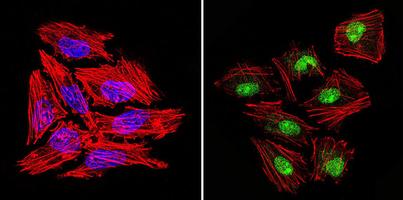
Immunofluorescent analysis of formalin-fixed, permeabilized HeLa cells, labeling GATA2 with ab173817 at 1/200 dilution (green). Cells were washed with PBST and incubated with a DyLight-conjugated secondary antibody. F-actin (red) was stained with a fluorescent phalloidin and nuclei (blue) were stained with DAPI.
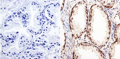
Immunohistochemical analysis of Human prostate tissue, labeling GATA2 with ab173817 at 1/100 dilution (right image). Detection was performed using a secondary antibody conjugated to HRP. DAB staining buffer was applied and tissues were counterstained with hematoxylin.
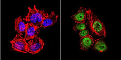
Immunofluorescent analysis of formalin-fixed, permeabilized U251 cells, labeling GATA2 with ab173817 at 1/20 dilution (green). Cells were washed with PBST and incubated with a DyLight-conjugated secondary antibody. F-actin (red) was stained with a fluorescent phalloidin and nuclei (blue) were stained with DAPI.
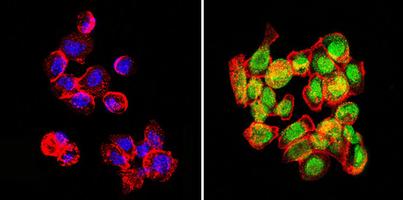
Immunofluorescent analysis of formalin-fixed, permeabilized Human cells, labeling GATA2 with ab173817 at 1/100 dilution (green). Cells were washed with PBST and incubated with a DyLight-conjugated secondary antibody. F-actin (red) was stained with a fluorescent phalloidin and nuclei (blue) were stained with DAPI.
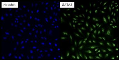
Immunofluorescent analysis of formalin-fixed, permeabilized HeLa cells, labeling GATA2 with ab173817 at 1/200 dilution (green). Cells were washed with PBS and incubated with a DyLight-488 conjugated secondary antibody. Nuclei (blue) were stained with Hoechst 33342 dye.

All lanes : Anti-GATA2 antibody - N-terminal (ab173817) at 1/500 dilutionLane 1 : mouse MEF cell lysateLane 2 : NTERRA cell lysateLysates/proteins at 25 µg per lane.Secondarygoat anti-rabbit-HRP at 1/20000 dilutiondeveloped using the ECL technique


