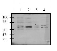
All lanes : Anti-Gata6 antibody (ab175349) at 1/1000 dilutionLane 1 : 293T cell lysateLane 2 : MCF7 cell lysateLane 3 : K562 cell lysateLane 4 : 3T3L1 cell lysateLysates/proteins at 25 µg per lane.SecondaryGoat anti-rabbit-HRP at 1/15000 dilutiondeveloped using the ECL technique

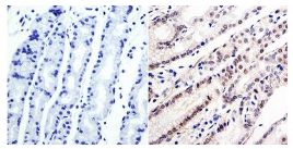
Immunohistochemical analysis of Human stomach tissue labeling Gata6 with ab175349 at 1/100 dilution. To expose target protein, antigen was retreived using 10mM sodium citrate followed by microwave treatment for 8-15 minutes. Endogenous peroxidases were blocked in 3% H202-methanol for 15 minutes and tissues were blocked in 3% BSA-PBS for 30 minutes at room temperature. Following labeling, tissues were washed in PBST and detection was performed using a secondary antibody conjugated to HRP. DAB staining buffer was applied and tissues were counterstained with hematoxylin and prepped for mounting.

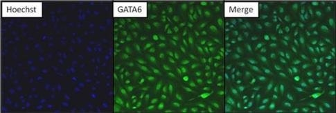
Immunofluorescent analysis of HeLa cells labeling Gata6 with ab175349 at 1/200 dilution (green). Formalin fixed cells were permeabilized with 0.1% Triton X-100 in TBS for 10 minutes at room temperature. Cells were then blocked with 5% normal goat serum for 15 minutes at room temperature. Following detection with the primary antibody, the cells were then washed with PBS and incubated with DyLight 488 goat-anti-rabbit secondary antibody at a dilution of 1/200 for 30 minutes at room temperature. Nuclei (blue) were stained with Hoechst 33342 dye.

