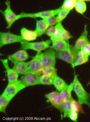
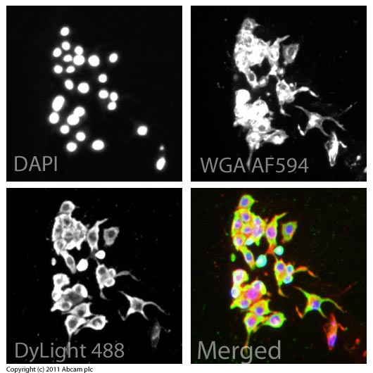
Anti-Gephyrin antibody
| Name | Anti-Gephyrin antibody |
|---|---|
| Supplier | Abcam |
| Catalog | ab32206 |
| Prices | $400.00 |
| Sizes | 100 µg |
| Host | Rabbit |
| Clonality | Polyclonal |
| Isotype | IgG |
| Applications | ICC/IF ICC/IF IHC-F WB |
| Species Reactivities | Mouse, Rat, Chicken, Human, Xenopus |
| Antigen | Synthetic peptide conjugated to KLH derived from within residues 700 to the C-terminus of Mouse Gephyrin |
| Blocking Peptide | Mouse Gephyrin peptide |
| Description | Rabbit Polyclonal |
| Gene | GPHN |
| Conjugate | Unconjugated |
| Supplier Page | Shop |
Product images
Product References
Disruption of centrifugal inhibition to olfactory bulb granule cells impairs - Disruption of centrifugal inhibition to olfactory bulb granule cells impairs
Nunez-Parra A, Maurer RK, Krahe K, Smith RS, Araneda RC. Proc Natl Acad Sci U S A. 2013 Sep 3;110(36):14777-82. doi:
Dynamic changes in neurexins' alternative splicing: role of Rho-associated - Dynamic changes in neurexins' alternative splicing: role of Rho-associated
Rozic G, Lupowitz Z, Piontkewitz Y, Zisapel N. PLoS One. 2011 Apr 12;6(4):e18579.
Morphological changes and synaptogenesis of corticothalamic neurons in the - Morphological changes and synaptogenesis of corticothalamic neurons in the
Hsu CI, Ho TS, Liou YR, Chang YC. Cereb Cortex. 2011 Apr;21(4):884-95.
Early developmental alterations in GABAergic protein expression in fragile X - Early developmental alterations in GABAergic protein expression in fragile X
Adusei DC, Pacey LK, Chen D, Hampson DR. Neuropharmacology. 2010 Sep;59(3):167-71.
High resolution in situ zymography reveals matrix metalloproteinase activity at - High resolution in situ zymography reveals matrix metalloproteinase activity at
Gawlak M, Gorkiewicz T, Gorlewicz A, Konopacki FA, Kaczmarek L, Wilczynski GM. Neuroscience. 2009 Jan 12;158(1):167-76.
Fluctuations in brain concentrations of neurosteroids are not associated to - Fluctuations in brain concentrations of neurosteroids are not associated to
Sassoe-Pognetto M, Follesa P, Panzanelli P, Perazzini AZ, Porcu P, Sogliano C, Cherchi C, Concas A. Brain Res. 2007 Sep 12;1169:1-8. Epub 2007 Jul 13.


