All lanes : Anti-Gephyrin antibody (ab25784) at 1 µg/mlLane 1 : Brain (Mouse) Tissue LysateLane 2 : Brain (Mouse) Tissue Lysate - normal tissue, 0 days old (ab7188)Lane 3 : NIH 3T3 (Mouse embryonic fibroblast cell line) Whole Cell Lysate (ab7179)Lane 4 : MEF1 (Mouse embryonic fibroblast cell line) Whole Cell LysateLane 5 : PC12 (Rat adrenal pheochromocytoma cell line) Whole Cell LysateLysates/proteins at 10 µg per lane.SecondaryGoat polyclonal to Rabbit IgG - H&L - Pre-Adsorbed (HRP) at 1/3000 dilutionPerformed under reducing conditions.

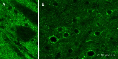
Immunohistochemistical detection of Gephyrin using antibody ab25784 on rat brain PFA perfusion-fixed free floating sections. Antigen retrieval step: None. Primary Antibody Dilution 1/300 incubated for 18 hours @ 20°C in PBS + 0.3 % Triton X100. Secondary Antibody: Goat anti-rabbit Alexa Fluor® 488 (1/1000). The submitted image is of a 30um rat brain section; a thinly punctuated staining was observed in the adult rat brain. The pictures show the staining obtained in the striatum (A) using the X20 objective and in the cerebral cortex using the X40 objective (B). The tissues were fixed with 4% PFA and later postfixed overnight in the same fixative. They were cryoprotected in 30% sucrose and cut using a cryostat.See Abreview

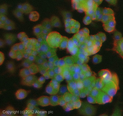
ICC/IF image of ab25784 stained PC12 cells. The cells were 4% formaldeyhde fixed (10 min) and then incubated in 1%BSA / 10% normal goat serum / 0.3M glycine in 0.1% PBS-Tween for 1h to permeabilise the cells and block non-specific protein-protein interactions. The cells were then incubated with the antibody (ab25784, 1µg/ml) overnight at +4°C. The secondary antibody (green) was ab96899, DyLight® 488 goat anti-rabbit IgG (H+L) used at a 1/250 dilution for 1h. Alexa Fluor® 594 WGA was used to label plasma membranes (red) at a 1/200 dilution for 1h. DAPI was used to stain the cell nuclei (blue) at a concentration of 1.43µM. This antibody also gave a positive result in 100% methanol fixed (5 min) XXX cells at 1µg/ml.


