Anti-Golgin 97 antibody
| Name | Anti-Golgin 97 antibody |
|---|---|
| Supplier | Abcam |
| Catalog | ab84340 |
| Prices | $382.00 |
| Sizes | 100 µg |
| Host | Rabbit |
| Clonality | Polyclonal |
| Isotype | IgG |
| Applications | ICC/IF ICC/IF IHC-P |
| Species Reactivities | Mouse, Human |
| Antigen | Synthetic peptide conjugated to KLH derived from within residues 750 to the C-terminus of Human Golgin 97 |
| Description | Rabbit Polyclonal |
| Gene | GOLGA1 |
| Conjugate | Unconjugated |
| Supplier Page | Shop |
Product images
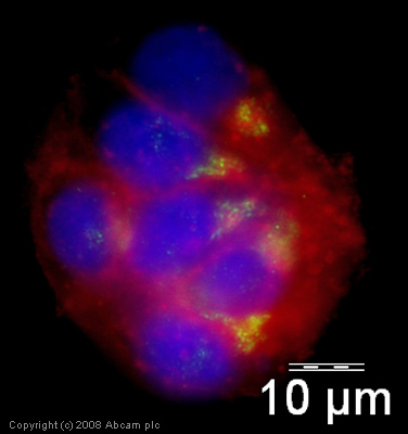 ICC/IF image of ab84340 stained human HepG2 cells. The cells were methanol fixed (5 min), permeabilised in 0.1% PBS-Tween (20 min) and incubated with the antibody (ab84340, 5µg/ml) for 1h at room temperature. 1%BSA / 10% normal goat serum / 0.3M glycine was used to block non-specific protein-protein interactions. The secondary antibody (green) was Alexa Fluor® 488 goat anti-rabbit IgG (H+L) used at a 1/1000 dilution for 1h. Alexa Fluor® 594 WGA was used to label plasma membranes (red). DAPI was used to stain the cell nuclei (blue).
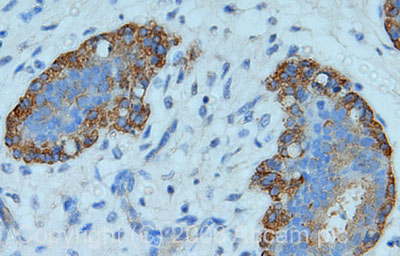 IHC image of Golgin 97 staining in human breast carcinoma FFPE section, performed on a BondTM system using the standard protocol F. The section was pre-treated using heat mediated antigen retrieval with sodium citrate buffer (pH6, epitope retrieval solution 1) for 20 mins. The section was then incubated with ab84340, 5µg/ml, for 8 mins at room temperature and detected using an HRP conjugated compact polymer system. DAB was used as the chromogen. The section was then counterstained with haematoxylin and mounted with DPX.
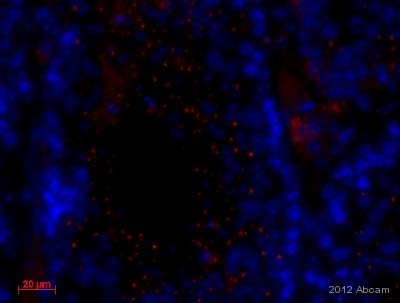 ab84340 staining Golgin 97 in murine testis tissue by Immunohistochemistry (Formalin/PFA-fixed paraffin-embedded sections).Tissue was fixed with formaldehyde and a heat mediated antigen retrieval step was performed using 0.01M sodium citrate. Samples were then blocked using 10%FCS, 0.5% BSA, 1% Triton X-100, 88.5% PBS X1 and then incubated with ab84340 at a 1/100 dilution for 10 hours at 4°C. The secondary used was an Alexa-Fluor 594 conjugated goat anti-rabbit IgG used at a 1/200 dilution.See Abreview
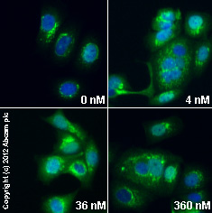 ab84340 staining Golgin-97 in MCF7 cells treated with brefeldin A (ab120299), by ICC/IF. Increase in Golgin-97 expression correlates with increased concentration of brefeldin A, as described in literature.The cells were incubated at 37øC for 1.5 h in media containing different concentrations of ab120299 (brefeldin A) in DMSO, fixed with 4% formaldehyde for 10 minutes at room temperature and blocked with PBS containing 10% goat serum, 0.3 M glycine, 1% BSA and 0.1% tween for 2h at room temperature. Staining of the treated cells with ab84340 (5 µg/ml) was performed overnight at 4øC in PBS containing 1% BSA and 0.1% tween. A DyLight 488 goat anti-rabbit polyclonal antibody (ab96899) at 1/250 dilution was used as the secondary antibody. Nuclei were counterstained with DAPI and are shown in blue.
Product References
Membrane adhesion dictates Golgi stacking and cisternal morphology. - Membrane adhesion dictates Golgi stacking and cisternal morphology.
Lee I, Tiwari N, Dunlop MH, Graham M, Liu X, Rothman JE. Proc Natl Acad Sci U S A. 2014 Feb 4;111(5):1849-54. doi:
Antibody-mediated inhibition of ricin toxin retrograde transport. - Antibody-mediated inhibition of ricin toxin retrograde transport.
Yermakova A, Klokk TI, Cole R, Sandvig K, Mantis NJ. MBio. 2014 Apr 8;5(2):e00995.
Fully synthetic polymer vesicles for intracellular delivery of antibodies in live - Fully synthetic polymer vesicles for intracellular delivery of antibodies in live
Canton I, Massignani M, Patikarnmonthon N, Chierico L, Robertson J, Renshaw SA, Warren NJ, Madsen JP, Armes SP, Lewis AL, Battaglia G. FASEB J. 2013 Jan;27(1):98-108.