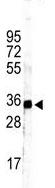
Anti-GRHPR antibody - C-terminal (ab176011) at 1/1000 dilution + Mouse kidney tissue lysate at 35 µg
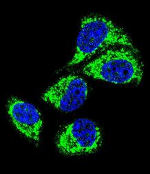
Immunofluorescence analysis of MCF-7 cells labeling GRHPR with ab176011 at a 1/10 dilution, followed by Alexa Fluor 488 conjugated goat anti-rabbit lgG. DAPI was used to stain the cell nuclear (blue).
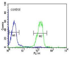
Flow cytometric analysis of HL60 cells (green) compared to a negative control cell (blue) using ab176011 at a 1/10 dilution. FITC-conjugated goat-anti-rabbit secondary antibodies were used for the analysis.

Immunohistochemical analysis of formalin fixed, paraffin embedded Human hepatocarcinoma tissue labeling GRHPR with ab176011 at a 1/50 dilution, followed by peroxidase conjugation of the secondary antibody and DAB staining.