Anti-HDAC3 antibody
| Name | Anti-HDAC3 antibody |
|---|---|
| Supplier | Abcam |
| Catalog | ab16047 |
| Host | Rabbit |
| Clonality | Polyclonal |
| Isotype | IgG |
| Applications | IHC-P ICC/IF ICC/IF ICC/IF WB IP |
| Species Reactivities | Mouse, Rat, Human, Deer, Zebrafish, Chicken, Xenopus |
| Antigen | Synthetic peptide conjugated to KLH derived from within residues 400 to the C-terminus of Human HDAC3 |
| Blocking Peptide | Human HDAC3 peptide |
| Description | Rabbit Polyclonal |
| Gene | HDAC3 |
| Conjugate | Unconjugated |
| Supplier Page | Shop |
Product images
 Lanes 1 & 6 : Anti-HDAC3 antibody (ab16047) at 1 µg/ml (Sample: HeLa nuclear lysate, 20 ug)Lanes 2 & 7 : Anti-HDAC3 antibody (ab16047) at 1 µg/ml (Sample: HeLa whole cell lysate, 20 ug)Lanes 3 & 8 : Anti-HDAC3 antibody (ab16047) at 1 µg/ml (Sample: A431 cell lysate, 20 ug)Lanes 4 & 9 : Anti-HDAC3 antibody (ab16047) at 1 µg/ml (Sample: Jurkat cell lysate, 20 ug)Lanes 5 & 10 : Anti-HDAC3 antibody (ab16047) at 1 µg/ml (Sample: HEK293 cell lysate, 20 ug)Lane 1 : As aboveLane 2 : As aboveLane 3 : As aboveLane 4 : As aboveLane 5 : As aboveLane 6 : Human HDAC3 peptide (ab16279) at 1 µg/mlLane 7 : Human HDAC3 peptide (ab16279) at 1 µg/mlLane 8 : Human HDAC3 peptide (ab16279) at 1 µg/mlLane 9 : Human HDAC3 peptide (ab16279) at 1 µg/mlLane 10 : Human HDAC3 peptide (ab16279) at 1 µg/mlSecondaryGoat Anti-Rabbit IgG H&L (HRP) (ab6721) at 1/5000 dilutionPerformed under reducing conditions.
 SK-N-SH cells were fixed in 4% paraformaldehyde, permeabilized in 0.5% Triton X-100 and incubated for 1 hour with ab16047 (1/100). The antibody clearly stained the nucleus (red). The cells were counterstained with DAPI (blue). 100x magnification.
 Interphase HeLa cells incubated with ab16047 (1/500). The antibody shows predominantly nuclear staining. The ab16047 staining is shown in green. The cells were counterstained with DAPI (red).
 ab16047 staining HDAC3 in human breast cancer tissue section by Immunohistochemistry (Formalin/PFA-fixed paraffin-embedded sections). Tissue underwent fixation in formaldehyde, heat mediated antigen retrieval in Citrate buffer pH 6.0 and blocking (5 minutes/peroxidase block then 10 minutes/protein block) for 15 minutes at 20°C. The primary antibody was diluted, 1/2000 and incubated with sample for 45 minutes at 20°C. A HRP conjugated goat polyclonal to rabbit IgG was used undiluted as secondary.See Abreview
 All lanes : Anti-HDAC3 antibody (ab16047) at 1 µg/mlLane 1 : Mouse 3T3 cell lysateLane 2 : Rat liver cell lysateLysates/proteins at 20 µg per lane.SecondaryGoat Anti-Rabbit IgG H&L (HRP) (ab6721) at 1/5000 dilutiondeveloped using the ECL techniquePerformed under reducing conditions.
![HDAC3 was immunoprecipitated using 0.5mg Hela whole cell extract, 5µg of Rabbit polyclonal to HDAC3 and 50µl of protein G magnetic beads (+). No antibody was added to the control (-). The antibody was incubated under agitation with Protein G beads for 10min, Hela whole cell extract lysate diluted in RIPA buffer was added to each sample and incubated for a further 10min under agitation.Proteins were eluted by addition of 40µl SDS loading buffer and incubated for 10min at 70oC; 10µl of each sample was separated on a SDS PAGE gel, transferred to a nitrocellulose membrane, blocked with 5% BSA and probed with ab16047.Secondary: Mouse monoclonal [SB62a] Secondary Antibody to Rabbit IgG light chain (HRP) (ab99697).Band: 49kDa: HDAC3.](http://www.bioprodhub.com/system/product_images/ab_products/2/sub_2/29387_HDAC3-Primary-antibodies-ab16047-17.jpg) HDAC3 was immunoprecipitated using 0.5mg Hela whole cell extract, 5µg of Rabbit polyclonal to HDAC3 and 50µl of protein G magnetic beads (+). No antibody was added to the control (-). The antibody was incubated under agitation with Protein G beads for 10min, Hela whole cell extract lysate diluted in RIPA buffer was added to each sample and incubated for a further 10min under agitation.Proteins were eluted by addition of 40µl SDS loading buffer and incubated for 10min at 70oC; 10µl of each sample was separated on a SDS PAGE gel, transferred to a nitrocellulose membrane, blocked with 5% BSA and probed with ab16047.Secondary: Mouse monoclonal [SB62a] Secondary Antibody to Rabbit IgG light chain (HRP) (ab99697).Band: 49kDa: HDAC3.
HDAC3 was immunoprecipitated using 0.5mg Hela whole cell extract, 5µg of Rabbit polyclonal to HDAC3 and 50µl of protein G magnetic beads (+). No antibody was added to the control (-). The antibody was incubated under agitation with Protein G beads for 10min, Hela whole cell extract lysate diluted in RIPA buffer was added to each sample and incubated for a further 10min under agitation.Proteins were eluted by addition of 40µl SDS loading buffer and incubated for 10min at 70oC; 10µl of each sample was separated on a SDS PAGE gel, transferred to a nitrocellulose membrane, blocked with 5% BSA and probed with ab16047.Secondary: Mouse monoclonal [SB62a] Secondary Antibody to Rabbit IgG light chain (HRP) (ab99697).Band: 49kDa: HDAC3.
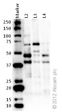 All lanes : Anti-HDAC3 antibody (ab16047) at 1 µg/mlLane 1 : MarkerLane 2 : Zebrafish brain homogenate (20ug)Lane 3 : Zebrafish heart homogenate (20ug)Lane 4 : Zebrafish liver homogenate (20ug)SecondaryDonkey polyclonal to Goat IgG – H&L – Pre-Adsorbed (HRP) at 1/6000 dilutiondeveloped using the ECL techniquePerformed under reducing conditions.
Product References
.
Monoaminergic and neuropeptidergic neurons have distinct expression profiles of - Monoaminergic and neuropeptidergic neurons have distinct expression profiles of
Takase K, Oda S, Kuroda M, Funato H. PLoS One. 2013;8(3):e58473.
.
Inhibition of histone deacetylase 3 causes replication stress in cutaneous T cell - Inhibition of histone deacetylase 3 causes replication stress in cutaneous T cell
Wells CE, Bhaskara S, Stengel KR, Zhao Y, Sirbu B, Chagot B, Cortez D, Khabele D, Chazin WJ, Cooper A, Jacques V, Rusche J, Eischen CM, McGirt LY, Hiebert SW. PLoS One. 2013 Jul 22;8(7):e68915.
HDAC3-dependent reversible lysine acetylation of cardiac myosin heavy chain - HDAC3-dependent reversible lysine acetylation of cardiac myosin heavy chain
Samant SA, Courson DS, Sundaresan NR, Pillai VB, Tan M, Zhao Y, Shroff SG, Rock RS, Gupta MP. J Biol Chem. 2011 Feb 18;286(7):5567-77.
Epigenetic regulation of a murine retrotransposon by a dual histone modification - Epigenetic regulation of a murine retrotransposon by a dual histone modification
Brunmeir R, Lagger S, Simboeck E, Sawicka A, Egger G, Hagelkruys A, Zhang Y, Matthias P, Miller WJ, Seiser C. PLoS Genet. 2010 Apr 29;6(4):e1000927.
Sp1 acetylation is associated with loss of DNA binding at promoters associated - Sp1 acetylation is associated with loss of DNA binding at promoters associated
Waby JS, Chirakkal H, Yu C, Griffiths GJ, Benson RS, Bingle CD, Corfe BM. Mol Cancer. 2010 Oct 15;9:275.
Deletion of histone deacetylase 3 reveals critical roles in S phase progression - Deletion of histone deacetylase 3 reveals critical roles in S phase progression
Bhaskara S, Chyla BJ, Amann JM, Knutson SK, Cortez D, Sun ZW, Hiebert SW. Mol Cell. 2008 Apr 11;30(1):61-72.
SYT-SSX1 and SYT-SSX2 interfere with repression of E-cadherin by snail and slug: - SYT-SSX1 and SYT-SSX2 interfere with repression of E-cadherin by snail and slug:
Saito T, Nagai M, Ladanyi M. Cancer Res. 2006 Jul 15;66(14):6919-27.