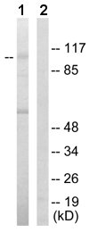
All lanes : Anti-Hexokinase 1 antibody (ab65069) at 1/500 dilutionLane 1 : extracts from HeLa cellsLane 2 : extracts from HeLa cells with immunizing peptide at 5 µgLysates/proteins at 5 µg per lane.
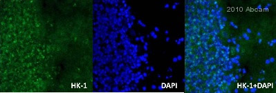
ab65069 staining Hexokinase 1 (green) in Rat brain tissue sections by Immunohistochemistry (IHC-Fr - frozen sections). Tissue was fixed with formaldehyde, permeabilized with 0.1% Triton X-100 and blocked with 5% BSA for 2 hours at 25°C. Samples were incubated with primary antibody (1/250 in PBS-T) for 2 hours at 25°C. An Alexa Fluor®488-conjugated Goat anti-rabbit IgG polyclonal (1/500) was used as the secondary antibody. Nuclei were stained by DAPI (blue).See Abreview
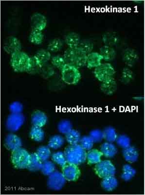
ab65069 staining Hexokinase 1 in murine bone marrow leukocytes by Immunocytochemistry/ Immunofluorescence. The cells were fixed in methanol and then blocked using 5% serum for 2 hours at 25°C. Samples were then incubated with the primary antibody at 1/250 for 16 hours at 4°C. The secondary antibody used was a goat anti-rabbit IgG conjugated to Alexa Fluor® 488 (green) used at a 1/500 dilution. DAPI was used for staining nuclei.See Abreview
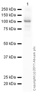
Anti-Hexokinase 1 antibody (ab65069) at 1/500 dilution + Active human Hexokinase 1 full length protein (ab85918) at 0.1 µgSecondaryGoat polyclonal to Rabbit IgG - H&L - Pre-Adsorbed (HRP) (ab65484) at 1/5000 dilutiondeveloped using the ECL techniquePerformed under reducing conditions.Exposure time : 4 minutes