Anti-HIF-1-alpha antibody [EP1215Y]
| Name | Anti-HIF-1-alpha antibody [EP1215Y] |
|---|---|
| Supplier | Abcam |
| Catalog | ab51608 |
| Prices | $403.00 |
| Sizes | 100 µl |
| Host | Rabbit |
| Clonality | Monoclonal |
| Isotype | IgG |
| Clone | EP1215Y |
| Applications | ICC/IF ICC/IF FC IP WB IHC-P |
| Species Reactivities | Mouse, Rat, Human |
| Antigen | Synthetic peptide (the amino acid sequence is considered to be commercially sensitive) corresponding to Human HIF-1-alpha aa 600-700 (C terminal) |
| Blocking Peptide | HIF-1-alpha peptide |
| Description | Rabbit Monoclonal |
| Gene | HIF1A |
| Conjugate | Unconjugated |
| Supplier Page | Shop |
Product images
Product References
Epithelial-mesenchymal transition induces endoplasmic-reticulum-stress response - Epithelial-mesenchymal transition induces endoplasmic-reticulum-stress response
Zeindl-Eberhart E, Brandl L, Liebmann S, Ormanns S, Scheel SK, Brabletz T, Kirchner T, Jung A. PLoS One. 2014 Jan 31;9(1):e87386.
Renal overexpression of atrial natriuretic peptide and hypoxia inducible - Renal overexpression of atrial natriuretic peptide and hypoxia inducible
Della Penna SL, Cao G, Carranza A, Zotta E, Gorzalczany S, Cerrudo CS, Rukavina Mikusic NL, Correa A, Trida V, Toblli JE, Roson MI, Fernandez BE. Biomed Res Int. 2014;2014:936978.
HIF-1 alpha overexpression correlates with poor overall survival and disease-free - HIF-1 alpha overexpression correlates with poor overall survival and disease-free
Chen L, Shi Y, Yuan J, Han Y, Qin R, Wu Q, Jia B, Wei B, Wei L, Dai G, Jiao S. PLoS One. 2014 Mar 10;9(3):e90678.
BAG3 and HIF-1 alpha coexpression detected by immunohistochemistry correlated - BAG3 and HIF-1 alpha coexpression detected by immunohistochemistry correlated
Xiao H, Tong R, Cheng S, Lv Z, Ding C, Du C, Xie H, Zhou L, Wu J, Zheng S. Biomed Res Int. 2014;2014:516518.
Mid- to late term hypoxia in the mouse alters placental morphology, - Mid- to late term hypoxia in the mouse alters placental morphology,
Cuffe JS, Walton SL, Singh RR, Spiers JG, Bielefeldt-Ohmann H, Wilkinson L, Little MH, Moritz KM. J Physiol. 2014 Jul 15;592(Pt 14):3127-41.
Divergent regulation of angiopoietin-1, angiopoietin-2, and vascular endothelial - Divergent regulation of angiopoietin-1, angiopoietin-2, and vascular endothelial
Tsuzuki T, Okada H, Cho H, Shimoi K, Miyashiro H, Yasuda K, Kanzaki H. Eur J Obstet Gynecol Reprod Biol. 2013 May;168(1):95-101. doi:
Ubc9 acetylation modulates distinct SUMO target modification and hypoxia - Ubc9 acetylation modulates distinct SUMO target modification and hypoxia
Hsieh YL, Kuo HY, Chang CC, Naik MT, Liao PH, Ho CC, Huang TC, Jeng JC, Hsu PH, Tsai MD, Huang TH, Shih HM. EMBO J. 2013 Mar 20;32(6):791-804.
Intratumor hypoxia promotes immune tolerance by inducing regulatory T cells via - Intratumor hypoxia promotes immune tolerance by inducing regulatory T cells via
Deng B, Zhu JM, Wang Y, Liu TT, Ding YB, Xiao WM, Lu GT, Bo P, Shen XZ. PLoS One. 2013 May 27;8(5):e63777.
Hypoxia-inducible factor-1alpha and MAPK co-regulate activation of hepatic - Hypoxia-inducible factor-1alpha and MAPK co-regulate activation of hepatic
Wang Y, Huang Y, Guan F, Xiao Y, Deng J, Chen H, Chen X, Li J, Huang H, Shi C. PLoS One. 2013 Sep 10;8(9):e74051.
Regulation of p53 by jagged1 contributes to angiotensin II-induced impairment of - Regulation of p53 by jagged1 contributes to angiotensin II-induced impairment of
Guan A, Gong H, Ye Y, Jia J, Zhang G, Li B, Yang C, Qian S, Sun A, Chen R, Ge J, Zou Y. PLoS One. 2013 Oct 3;8(10):e76529.
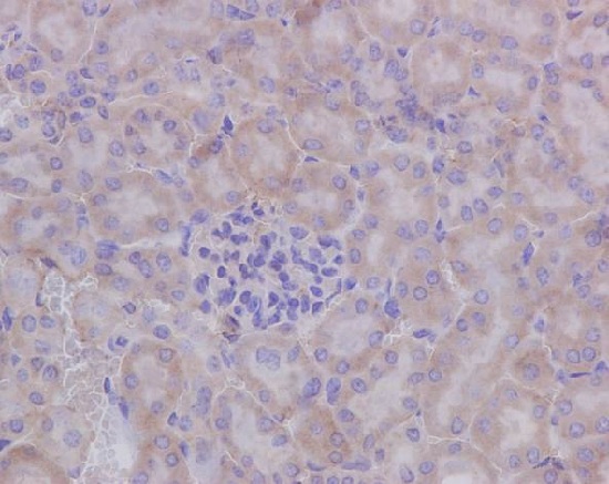
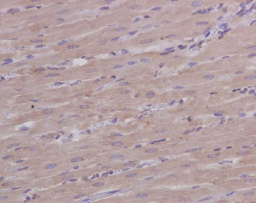
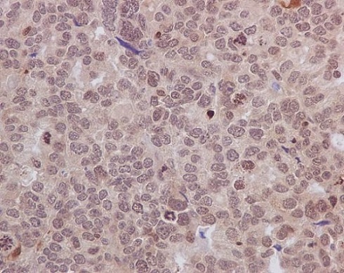
![Anti-HIF-1-alpha antibody [EP1215Y] (ab51608) at 1/100 dilution + Ramos Cells treated with Cocl2 at 10 µgSecondaryGoat Anti-Rabbit IgG, (H+L), HRP- conjugated at 1/1000 dilution](http://www.bioprodhub.com/system/product_images/ab_products/2/sub_3/549_ab51608-239742-51608-WB.jpg)
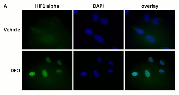
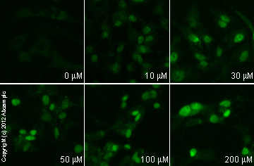
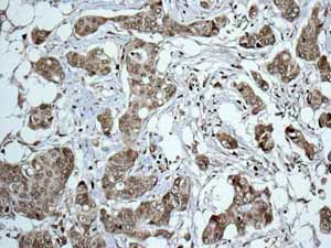
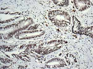
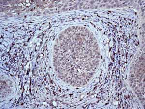
![All lanes : Anti-HIF-1-alpha antibody [EP1215Y] (ab51608) at 1/2000 dilution (Unpurified)Lane 1 : HeLa (Human epithelial carcinoma cell line) Nuclear Lysate (ab150036)Lane 2 : Hela-DFO treated (0.5mM, 24h) Nuclear Lysate (ab180880)Lysates/proteins at 40 µg per lane.SecondaryGoat Anti-Rabbit IgG H&L (HRP) (ab97051) at 1/10000 dilutiondeveloped using the ECL techniquePerformed under reducing conditions.](http://www.bioprodhub.com/system/product_images/ab_products/2/sub_3/555_ab51608-210519-WBYI010605C23.jpg)
![HIF-1-alpha was immunoprecipitated using 0.5mg HeLa Nuclear DFO treated whole cell extract (ab180880), 5µg of Rabbit polyclonal to HIF1 alpha and 50µl of protein G magnetic beads (+). No antibody was added to the control (-).The antibody was incubated under agitation with Protein G beads for 10min, HeLa DFO treated whole cell extract lysate diluted in RIPA buffer was added to each sample and incubated for a further 10min under agitation.Proteins were eluted by addition of 40µl SDS loading buffer and incubated for 10min at 70°C; 10µl of each sample was separated on a SDS PAGE gel, transferred to a nitrocellulose membrane, blocked with 5% BSA and probed with unpurified ab51608.Secondary: Mouse monoclonal [SB62a] Secondary Antibody to Rabbit IgG light chain (HRP) (ab99697).Band: 110kDa; HIF1 alpha](http://www.bioprodhub.com/system/product_images/ab_products/2/sub_3/556_ab51608-209834-IPVI004ab5160812m.jpg)
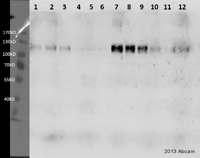
![All lanes : Anti-HIF-1-alpha antibody [EP1215Y] (ab51608) at 1/2000 dilution (unpurified)Lane 1 : HeLa Whole Cell Lysate (untreated, negative control)Lane 2 : HeLa DFO treated (0.5mM, 24h) Whole Cell LysateLane 3 : HeLa Nuclear Cell Lysate (untreated, negative control)Lane 4 : HeLa Nuclear DFO treated (0.5mM, 24h) Cell LysateLysates/proteins at 40 µg per lane.SecondaryGoat Anti-Rabbit IgG H&L (HRP) (ab97051) at 1/10000 dilutionPerformed under reducing conditions.](http://www.bioprodhub.com/system/product_images/ab_products/2/sub_3/558_ab51608-204431-WBYI010605021.jpg)
