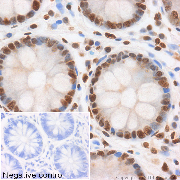
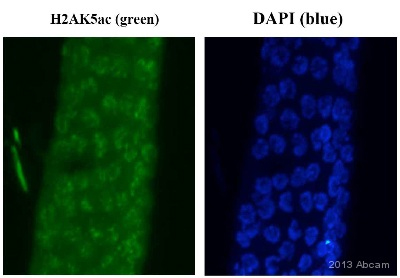
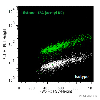
Anti-Histone H2A (acetyl K5) antibody - ChIP Grade
| Name | Anti-Histone H2A (acetyl K5) antibody - ChIP Grade |
|---|---|
| Supplier | Abcam |
| Catalog | ab1764 |
| Prices | $401.00 |
| Sizes | 100 µl |
| Host | Rabbit |
| Clonality | Polyclonal |
| Isotype | IgG |
| Applications | ChIP ELISA IP WB IHC-P ICC/IF ICC/IF |
| Species Reactivities | Human, C. elegans, Drosophila, Mammalian |
| Antigen | Synthetic peptide: SGRGKAcQGGKYC conjugated to Ovalbumin |
| Description | Rabbit Polyclonal |
| Gene | CELE_ZK1251.1 |
| Conjugate | Unconjugated |
| Supplier Page | Shop |
Product images
Product References
SIRT6 is a histone H3 lysine 9 deacetylase that modulates telomeric chromatin. - SIRT6 is a histone H3 lysine 9 deacetylase that modulates telomeric chromatin.
Michishita E, McCord RA, Berber E, Kioi M, Padilla-Nash H, Damian M, Cheung P, Kusumoto R, Kawahara TL, Barrett JC, Chang HY, Bohr VA, Ried T, Gozani O, Chua KF. Nature. 2008 Mar 27;452(7186):492-6.
Mrg15 null and heterozygous mouse embryonic fibroblasts exhibit DNA-repair - Mrg15 null and heterozygous mouse embryonic fibroblasts exhibit DNA-repair
Garcia SN, Kirtane BM, Podlutsky AJ, Pereira-Smith OM, Tominaga K. FEBS Lett. 2007 Nov 13;581(27):5275-81. Epub 2007 Oct 18.
Np95 is implicated in pericentromeric heterochromatin replication and in major - Np95 is implicated in pericentromeric heterochromatin replication and in major
Papait R, Pistore C, Negri D, Pecoraro D, Cantarini L, Bonapace IM. Mol Biol Cell. 2007 Mar;18(3):1098-106. Epub 2006 Dec 20.
Myc-binding-site recognition in the human genome is determined by chromatin - Myc-binding-site recognition in the human genome is determined by chromatin
Guccione E, Martinato F, Finocchiaro G, Luzi L, Tizzoni L, Dall' Olio V, Zardo G, Nervi C, Bernard L, Amati B. Nat Cell Biol. 2006 Jul;8(7):764-70. Epub 2006 Jun 11.
Transcription-induced chromatin remodeling at the c-myc gene involves the local - Transcription-induced chromatin remodeling at the c-myc gene involves the local
Farris SD, Rubio ED, Moon JJ, Gombert WM, Nelson BH, Krumm A. J Biol Chem. 2005 Jul 1;280(26):25298-303. Epub 2005 May 6.


