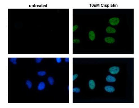
ab184520 detects H2A.X protein at nuclear by immunofluorescent analysis. Sample: 10uM Cisplatin treated (right) or untreated (left) HeLa cells were fixed in 4% paraformaldehyde for 15 min. Green: H2A.X protein stained with ab184520 diluted at 1:500. Blue: Hoechst 33342 staining. Bottom panels show merged images

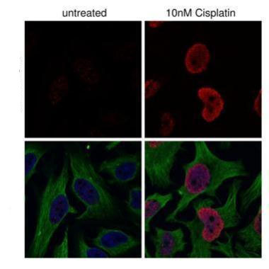
ab184520 detects H2A.X protein at nuclear by confocal immunofluorescent analysis.Sample: 10nM Cisplatin treated (right) or untreated (left) HeLa cells were fixed in 4% paraformaldehyde for 15 min.Red: H2A.X protein stained with ab184520 diluted at 1:500.Green: alpha Tubulin antibody diluted at 1:1000.Blue: Hoechst 33342 staining. Bottom panels show merged images.
![All lanes : Anti-Histone H2A.X (phospho S139) antibody [GT2311] (ab184520) at 1/1000 dilutionLane 1 : HCT116 whole cell lysate (untreated)Lane 2 : HCT116 whole cell lysate (30 µM cisplatin treatment for 24hr)Lysates/proteins at 30 µg per lane.](http://www.bioprodhub.com/system/product_images/ab_products/2/sub_3/1163_ab184520-215123-WB1.jpg)
All lanes : Anti-Histone H2A.X (phospho S139) antibody [GT2311] (ab184520) at 1/1000 dilutionLane 1 : HCT116 whole cell lysate (untreated)Lane 2 : HCT116 whole cell lysate (30 µM cisplatin treatment for 24hr)Lysates/proteins at 30 µg per lane.
![All lanes : Anti-Histone H2A.X (phospho S139) antibody [GT2311] (ab184520) at 1/1000 dilutionLane 1 : NIH-3T3 whole cell lysate(untreated)Lane 2 : NIH-3T3 whole cell lysate (30 µM cisplatin treatment for 24hr)Lysates/proteins at 30 µg per lane.](http://www.bioprodhub.com/system/product_images/ab_products/2/sub_3/1164_ab184520-215122-WB1.jpg)
All lanes : Anti-Histone H2A.X (phospho S139) antibody [GT2311] (ab184520) at 1/1000 dilutionLane 1 : NIH-3T3 whole cell lysate(untreated)Lane 2 : NIH-3T3 whole cell lysate (30 µM cisplatin treatment for 24hr)Lysates/proteins at 30 µg per lane.

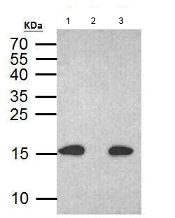
ab184520 immunoprecipitates histone H2A.X (phospho S139) protein in IP at 2 μg.1. 500 μg HCT116 with CPT 30 μM treatment 24 hr whole cell lysate/extract.2. 30 μg HCT116 whole with CPT 30 uM treatment cell lysate/extract .3. Control with 2 μg of preimmune mouse IgG C. The immunoprecipitated histone H2A.X (phospho S139) protein was detected by ab184520 diluted at 1:1000. 15% SDS-PAGE. Anti-mouse IgG was used as a secondary reagent.


![All lanes : Anti-Histone H2A.X (phospho S139) antibody [GT2311] (ab184520) at 1/1000 dilutionLane 1 : HCT116 whole cell lysate (untreated)Lane 2 : HCT116 whole cell lysate (30 µM cisplatin treatment for 24hr)Lysates/proteins at 30 µg per lane.](http://www.bioprodhub.com/system/product_images/ab_products/2/sub_3/1163_ab184520-215123-WB1.jpg)
![All lanes : Anti-Histone H2A.X (phospho S139) antibody [GT2311] (ab184520) at 1/1000 dilutionLane 1 : NIH-3T3 whole cell lysate(untreated)Lane 2 : NIH-3T3 whole cell lysate (30 µM cisplatin treatment for 24hr)Lysates/proteins at 30 µg per lane.](http://www.bioprodhub.com/system/product_images/ab_products/2/sub_3/1164_ab184520-215122-WB1.jpg)
