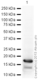
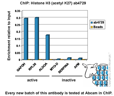
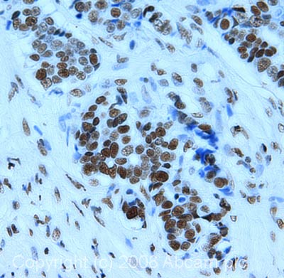
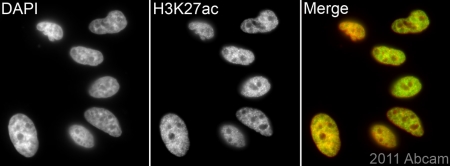
Anti-Histone H3 (acetyl K27) antibody - ChIP Grade
| Name | Anti-Histone H3 (acetyl K27) antibody - ChIP Grade |
|---|---|
| Supplier | Abcam |
| Catalog | ab4729 |
| Prices | $404.00 |
| Sizes | 100 µg |
| Host | Rabbit |
| Clonality | Polyclonal |
| Isotype | IgG |
| Applications | IHC-F ICC/IF ICC/IF WB IHC-P ChIPseq ChIP ChIP ChIP Array |
| Species Reactivities | Mouse, Rat, Chicken, Bovine, Human, Arabidopsis thaliana, Drosophila, Monkey, Plasmodium, Rice, Xenopus |
| Antigen | Synthetic peptide conjugated to KLH derived from within residues 1 - 100 of Human Histone H3, acetylated at K27 |
| Description | Rabbit Polyclonal |
| Gene | HIST1H3A |
| Conjugate | Unconjugated |
| Supplier Page | Shop |
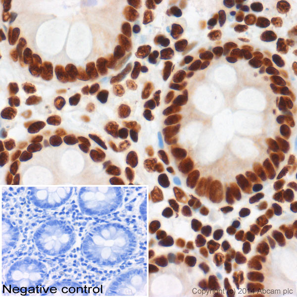
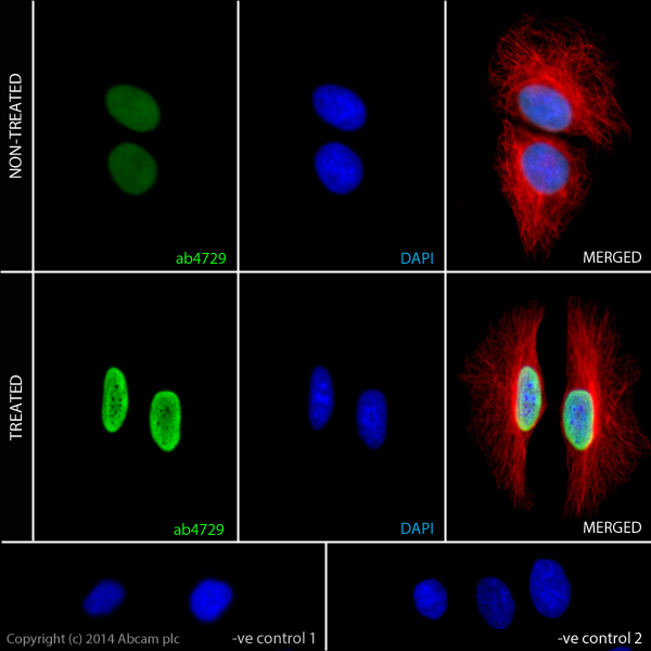
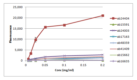
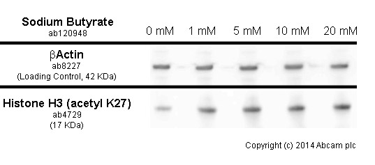
Product images
Product References
NOTCH1-RBPJ complexes drive target gene expression through dynamic interactions - NOTCH1-RBPJ complexes drive target gene expression through dynamic interactions
Wang H, Zang C, Taing L, Arnett KL, Wong YJ, Pear WS, Blacklow SC, Liu XS, Aster JC. Proc Natl Acad Sci U S A. 2014 Jan 14;111(2):705-10. doi:
Analysis of chromatin-state plasticity identifies cell-type-specific regulators - Analysis of chromatin-state plasticity identifies cell-type-specific regulators
Pinello L, Xu J, Orkin SH, Yuan GC. Proc Natl Acad Sci U S A. 2014 Jan 21;111(3):E344-53. doi:
The hematopoietic regulator TAL1 is required for chromatin looping between the - The hematopoietic regulator TAL1 is required for chromatin looping between the
Yun WJ, Kim YW, Kang Y, Lee J, Dean A, Kim A. Nucleic Acids Res. 2014 Apr;42(7):4283-93.
LSD1 regulates pluripotency of embryonic stem/carcinoma cells through histone - LSD1 regulates pluripotency of embryonic stem/carcinoma cells through histone
Yin F, Lan R, Zhang X, Zhu L, Chen F, Xu Z, Liu Y, Ye T, Sun H, Lu F, Zhang H. Mol Cell Biol. 2014 Jan;34(2):158-79.
Targeted chromatin profiling reveals novel enhancers in Ig H and Ig L chain Loci. - Targeted chromatin profiling reveals novel enhancers in Ig H and Ig L chain Loci.
Predeus AV, Gopalakrishnan S, Huang Y, Tang J, Feeney AJ, Oltz EM, Artyomov MN. J Immunol. 2014 Feb 1;192(3):1064-70.
A single allele of Hdac2 but not Hdac1 is sufficient for normal mouse brain - A single allele of Hdac2 but not Hdac1 is sufficient for normal mouse brain
Hagelkruys A, Lagger S, Krahmer J, Leopoldi A, Artaker M, Pusch O, Zezula J, Weissmann S, Xie Y, Schofer C, Schlederer M, Brosch G, Matthias P, Selfridge J, Lassmann H, Knoblich JA, Seiser C. Development. 2014 Feb;141(3):604-16.
Inhibition of endothelial ERK signalling by Smad1/5 is essential for - Inhibition of endothelial ERK signalling by Smad1/5 is essential for
Zhang C, Lv J, He Q, Wang S, Gao Y, Meng A, Yang X, Liu F. Nat Commun. 2014 Mar 11;5:3431.
beta-agonists selectively modulate proinflammatory gene expression in skeletal - beta-agonists selectively modulate proinflammatory gene expression in skeletal
Kolmus K, Van Troys M, Van Wesemael K, Ampe C, Haegeman G, Tavernier J, Gerlo S. PLoS One. 2014 Mar 6;9(6):e90649.
Combined protein- and nucleic acid-level effects of rs1143679 (R77H), a - Combined protein- and nucleic acid-level effects of rs1143679 (R77H), a
Maiti AK, Kim-Howard X, Motghare P, Pradhan V, Chua KH, Sun C, Arango-Guerrero MT, Ghosh K, Niewold TB, Harley JB, Anaya JM, Looger LL, Nath SK. Hum Mol Genet. 2014 Aug 1;23(15):4161-76.
Acetylation of histone H3 at lysine 64 regulates nucleosome dynamics and - Acetylation of histone H3 at lysine 64 regulates nucleosome dynamics and
Di Cerbo V, Mohn F, Ryan DP, Montellier E, Kacem S, Tropberger P, Kallis E, Holzner M, Hoerner L, Feldmann A, Richter FM, Bannister AJ, Mittler G, Michaelis J, Khochbin S, Feil R, Schuebeler D, Owen-Hughes T, Daujat S, Schneider R. Elife. 2014 Mar 25;3:e01632.
