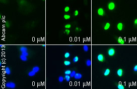
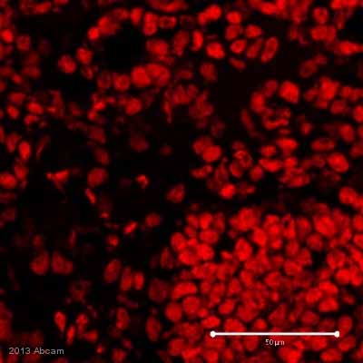
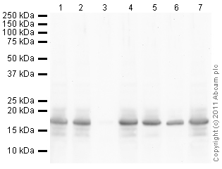
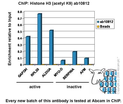
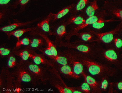
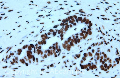
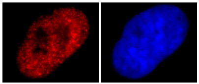
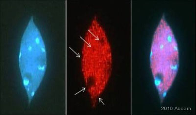
Anti-Histone H3 (acetyl K9) antibody - ChIP Grade
| Name | Anti-Histone H3 (acetyl K9) antibody - ChIP Grade |
|---|---|
| Supplier | Abcam |
| Catalog | ab10812 |
| Prices | $403.00 |
| Sizes | 100 µg |
| Host | Rabbit |
| Clonality | Polyclonal |
| Isotype | IgG |
| Applications | IHC-F IHC-P ChIP IP WB ChIPseq ICC/IF ICC/IF FC |
| Species Reactivities | Mouse, Rat, Bovine, Human, Yeast, Arabidopsis thaliana, C. elegans, Drosophila, Zebrafish, Marmoset, Rice, Broad species |
| Antigen | Synthetic peptide conjugated to KLH derived from within residues 1 - 100 of Human Histone H3, acetylated at K9 |
| Blocking Peptide | Human Histone H3 (acetyl K9) peptide |
| Description | Rabbit Polyclonal |
| Gene | HIST1H3A |
| Conjugate | Unconjugated |
| Supplier Page | Shop |
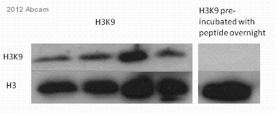
Product images
Product References
Glycogen synthase kinase (GSK) 3beta phosphorylates and protects nuclear myosin - Glycogen synthase kinase (GSK) 3beta phosphorylates and protects nuclear myosin
Sarshad AA, Corcoran M, Al-Muzzaini B, Borgonovo-Brandter L, Von Euler A, Lamont D, Visa N, Percipalle P. PLoS Genet. 2014 Jun 5;10(6):e1004390.
Differential acetylation of histone H3 at the regulatory region of OsDREB1b - Differential acetylation of histone H3 at the regulatory region of OsDREB1b
Roy D, Paul A, Roy A, Ghosh R, Ganguly P, Chaudhuri S. PLoS One. 2014 Jun 18;9(6):e100343.
T cell activation regulates CD6 alternative splicing by transcription dynamics - T cell activation regulates CD6 alternative splicing by transcription dynamics
da Gloria VG, Martins de Araujo M, Mafalda Santos A, Leal R, de Almeida SF, Carmo AM, Moreira A. J Immunol. 2014 Jul 1;193(1):391-9.
.
Inhibition of DNA methylation alters chromatin organization, nuclear positioning - Inhibition of DNA methylation alters chromatin organization, nuclear positioning
Bockor VV, Barisic D, Horvat T, Maglica Z, Vojta A, Zoldos V. PLoS One. 2014 Aug 5;9(8):e103954.
A robust chromatin immunoprecipitation protocol for studying transcription - A robust chromatin immunoprecipitation protocol for studying transcription
Li W, Lin YC, Li Q, Shi R, Lin CY, Chen H, Chuang L, Qu GZ, Sederoff RR, Chiang VL. Nat Protoc. 2014 Sep;9(9):2180-93.
Pleckstrin homology domain leucine-rich repeat protein phosphatases set the - Pleckstrin homology domain leucine-rich repeat protein phosphatases set the
Reyes G, Niederst M, Cohen-Katsenelson K, Stender JD, Kunkel MT, Chen M, Brognard J, Sierecki E, Gao T, Nowak DG, Trotman LC, Glass CK, Newton AC. Proc Natl Acad Sci U S A. 2014 Sep 23;111(38):E3957-65. doi:
Alcohol consumption during gestation causes histone3 lysine9 hyperacetylation and - Alcohol consumption during gestation causes histone3 lysine9 hyperacetylation and
Pan B, Zhu J, Lv T, Sun H, Huang X, Tian J. Alcohol Clin Exp Res. 2014 Sep;38(9):2396-402.
Dose- and time-dependent epigenetic changes in the livers of Fisher 344 rats - Dose- and time-dependent epigenetic changes in the livers of Fisher 344 rats
Conti Ad, Kobets T, Escudero-Lourdes C, Montgomery B, Tryndyak V, Beland FA, Doerge DR, Pogribny IP. Toxicol Sci. 2014 Jun;139(2):371-80.
Active, phosphorylated fingolimod inhibits histone deacetylases and facilitates - Active, phosphorylated fingolimod inhibits histone deacetylases and facilitates
Hait NC, Wise LE, Allegood JC, O'Brien M, Avni D, Reeves TM, Knapp PE, Lu J, Luo C, Miles MF, Milstien S, Lichtman AH, Spiegel S. Nat Neurosci. 2014 Jul;17(7):971-80.
