Anti-Histone H3 (phospho T32) antibody
| Name | Anti-Histone H3 (phospho T32) antibody |
|---|---|
| Supplier | Abcam |
| Catalog | ab4076 |
| Prices | $401.00 |
| Sizes | 100 µg |
| Host | Rabbit |
| Clonality | Polyclonal |
| Isotype | IgG |
| Applications | ICC/IF ICC/IF IHC-P WB ICC/IF |
| Species Reactivities | Human, Arabidopsis thaliana, Mouse, Rat, Chicken, Bovine, Xenopus, Drosophila, Zebrafish |
| Antigen | Synthetic peptide corresponding to Human Histone H3 aa 1-100 (phospho T32) conjugated to Keyhole Limpet Haemocyanin (KLH) |
| Blocking Peptide | Human Histone H3 (phospho T32) peptide |
| Description | Rabbit Polyclonal |
| Gene | H3F3A |
| Conjugate | Unconjugated |
| Supplier Page | Shop |
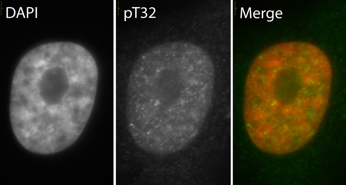
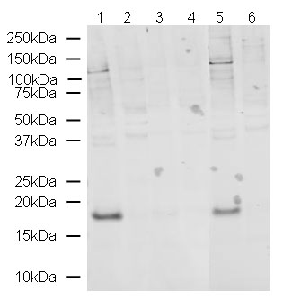
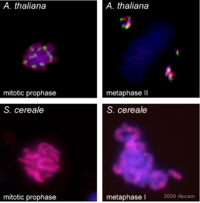
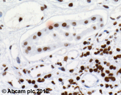
Product images
Product References
beta-N-Acetylglucosamine (O-GlcNAc) is a novel regulator of mitosis-specific - beta-N-Acetylglucosamine (O-GlcNAc) is a novel regulator of mitosis-specific
Fong JJ, Nguyen BL, Bridger R, Medrano EE, Wells L, Pan S, Sifers RN. J Biol Chem. 2012 Apr 6;287(15):12195-203.
Distribution patterns of phosphorylated Thr 3 and Thr 32 of histone H3 in plant - Distribution patterns of phosphorylated Thr 3 and Thr 32 of histone H3 in plant
Caperta AD, Rosa M, Delgado M, Karimi R, Demidov D, Viegas W, Houben A. Cytogenet Genome Res. 2008;122(1):73-9.
Distribution patterns of phosphorylated Thr 3 and Thr 32 of histone H3 in plant - Distribution patterns of phosphorylated Thr 3 and Thr 32 of histone H3 in plant
Caperta AD, Rosa M, Delgado M, Karimi R, Demidov D, Viegas W, Houben A. Cytogenet Genome Res. 2008;122(1):73-9.
Chromatin decondensation and nuclear reprogramming by nucleoplasmin. - Chromatin decondensation and nuclear reprogramming by nucleoplasmin.
Tamada H, Van Thuan N, Reed P, Nelson D, Katoku-Kikyo N, Wudel J, Wakayama T, Kikyo N. Mol Cell Biol. 2006 Feb;26(4):1259-71.


