![ab177068 staining Histone H4 (formyl K12) in HeLa cells. The cells were fixed with 100% methanol (5min) and then permeabilised using 0.1% PBS-Triton. Cells were then blocked in 1% BSA/10% normal goat serum/0.3M glycine in 0.1%PBS-Tween for 1h. The cells were then incubated with ab177068 at 1μg/ml overnight at +4°C, followed by a further incubation at room temperature for 1h with an AlexaFluor®488 Goat anti-Rabbit secondary (ab150081) at 1/1000 dilution (shown in green). AlexaFluor®594 Mouse monoclonal [DM1A] to alpha Tubulin (ab195889) - Microtubule Marker was used at a 1/200 dilution and incubated for 1h with the cells, to label Microtubules (shown in Red). The nuclear counter stain is DAPI (blue), which was added to the secondary antibody mixture.](http://www.bioprodhub.com/system/product_images/ab_products/2/sub_3/2307_ab177068-240656-PrimConjICCIFdsimagete.jpg)
ab177068 staining Histone H4 (formyl K12) in HeLa cells. The cells were fixed with 100% methanol (5min) and then permeabilised using 0.1% PBS-Triton. Cells were then blocked in 1% BSA/10% normal goat serum/0.3M glycine in 0.1%PBS-Tween for 1h. The cells were then incubated with ab177068 at 1μg/ml overnight at +4°C, followed by a further incubation at room temperature for 1h with an AlexaFluor®488 Goat anti-Rabbit secondary (ab150081) at 1/1000 dilution (shown in green). AlexaFluor®594 Mouse monoclonal [DM1A] to alpha Tubulin (ab195889) - Microtubule Marker was used at a 1/200 dilution and incubated for 1h with the cells, to label Microtubules (shown in Red). The nuclear counter stain is DAPI (blue), which was added to the secondary antibody mixture.

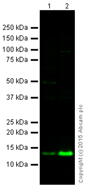
All lanes : Anti-Histone H4 (formyl K12) antibody (ab177068) at 1 µg/mlLane 1 : HeLa (Human epithelial carcinoma cell line) Whole Cell LysateLane 2 : HeLa Nuclear Prep (0.5% Triton X-100 insoluble fraction)Lysates/proteins at 10 µg per lane.SecondaryGoat Anti-Rabbit IgG H&L (Alexa Fluor® 790) at 1/10000 dilution

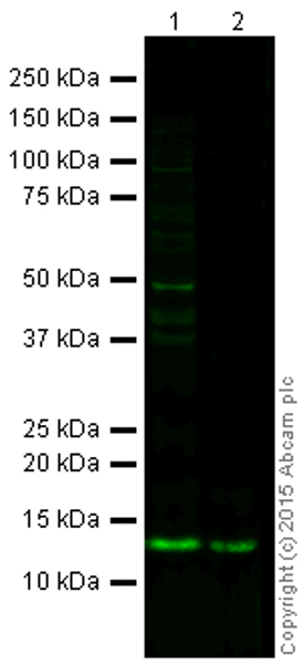
All lanes : Anti-Histone H4 (formyl K12) antibody (ab177068) at 1 µg/mlLane 1 : NIH 3T3 (Mouse embryonic fibroblast cell line) Whole Cell Lysate at 10 µgLane 2 : Calf Thymus Histone Preparation Nuclear Lysate at 0.5 µgSecondaryGoat Anti-Rabbit IgG H&L (Alexa Fluor® 790) at 1/10000 dilution
![ab177068 staining Histone H4 (formyl K12) in HeLa cells. The cells were fixed with 100% methanol (5min) and then permeabilised using 0.1% PBS-Triton. Cells were then blocked in 1% BSA/10% normal goat serum/0.3M glycine in 0.1%PBS-Tween for 1h. The cells were then incubated with ab177068 at 1μg/ml overnight at +4°C, followed by a further incubation at room temperature for 1h with an AlexaFluor®488 Goat anti-Rabbit secondary (ab150081) at 1/1000 dilution (shown in green). AlexaFluor®594 Mouse monoclonal [DM1A] to alpha Tubulin (ab195889) - Microtubule Marker was used at a 1/200 dilution and incubated for 1h with the cells, to label Microtubules (shown in Red). The nuclear counter stain is DAPI (blue), which was added to the secondary antibody mixture.](http://www.bioprodhub.com/system/product_images/ab_products/2/sub_3/2307_ab177068-240656-PrimConjICCIFdsimagete.jpg)

