Anti-HMGCS1 antibody
| Name | Anti-HMGCS1 antibody |
|---|---|
| Supplier | Abcam |
| Catalog | ab87246 |
| Prices | $384.00 |
| Sizes | 50 µg |
| Host | Rabbit |
| Clonality | Polyclonal |
| Isotype | IgG |
| Applications | WB ICC/IF ICC/IF IHC-P |
| Species Reactivities | Human, Mouse, Rat, Rabbit, Horse, Guinea Pig, Bovine, Cat, Dog, Pig |
| Antigen | Synthetic peptide corresponding to a region within the internal sequence amino acids 252-301 (DFTLNDFGFM IFHSPYCKLV QKSLARMLLN DFLNDQNRDK NSIYSGLEAF) of Human HMGCS1, NP_002121 Run BLAST with Run BLAST with |
| Description | Rabbit Polyclonal |
| Gene | HMGCS1 |
| Conjugate | Unconjugated |
| Supplier Page | Shop |
Product images
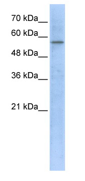 Anti-HMGCS1 antibody (ab87246) at 1 µg/ml + transfected 293T cell lysate at 10 µgSecondaryHRP conjugated anti-Rabbit IgG at 1/50000 dilution
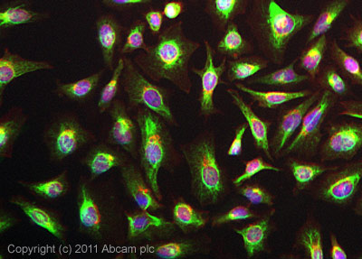 ICC/IF image of ab87246 stained HeLa cells. The cells were 4% formaldehyde fixed (10 min) and then incubated in 1%BSA / 10% normal goat serum / 0.3M glycine in 0.1% PBS-Tween for 1h to permeabilise the cells and block non-specific protein-protein interactions. The cells were then incubated with the antibody (ab87246, 5µg/ml) overnight at +4°C. The secondary antibody (green) was ab96899, DyLight® 488 goat anti-rabbit IgG (H+L) used at a 1/250 dilution for 1h.Alexa Fluor® 594 WGA was used to label plasma membranes (red) at a 1/200 dilution for 1h. DAPI was used to stain the cell nuclei (blue) at a concentration of 1.43µM.
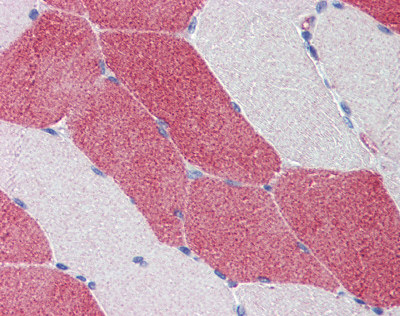 ab87246 staining HMGCS1 at 5 µg/ml in Human skeletal muscle tissue by Immunohistochemistry (Formalin/PFA-fixed paraffin-embedded sections).