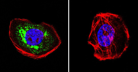
Immunocytochemistry/Immunofluorescence analysis of HSC70 Interacting Protein HIP in A431 cells. Cells were grown on chamber slides and fixed with formaldehyde prior to staining. Cells were probed without (control) or with ab2813 (1:100) overnight at 4°C, washed with PBS and incubated with a DyLight-488 conjugated secondary antibody. HSC70 Interacting Protein HIP staining (green), F-Actin staining with Phalloidin (red) and nuclei with DAPI (blue) is shown. Images were taken at 60X magnification.

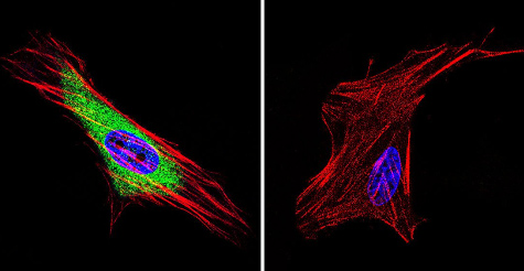
Immunocytochemistry/Immunofluorescence analysis of HSC70 Interacting Protein HIP in HeLa cells. Cells were grown on chamber slides and fixed with formaldehyde prior to staining. Cells were probed without (control) or with ab2813 (1:20) overnight at 4°C, washed with PBS and incubated with a DyLight-488 conjugated secondary antibody. HSC70 Interacting Protein HIP staining (green), F-Actin staining with Phalloidin (red) and nuclei with DAPI (blue) is shown. Images were taken at 60X magnification.

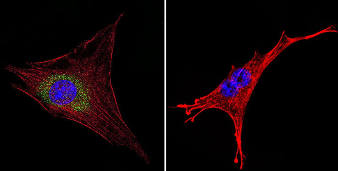
Immunocytochemistry/Immunofluorescence analysis of HSC70 Interacting Protein HIP in NIH-3T3 cells. Cells were grown on chamber slides and fixed with formaldehyde prior to staining. Cells were probed without (control) or with ab2813 (1:200) overnight at 4°C, washed with PBS and incubated with a DyLight-488 conjugated secondary antibody. HSC70 Interacting Protein HIP staining (green), F-Actin staining with Phalloidin (red) and nuclei with DAPI (blue) is shown. Images were taken at 60X magnification.


