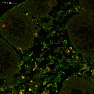
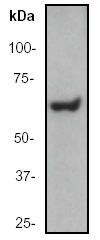
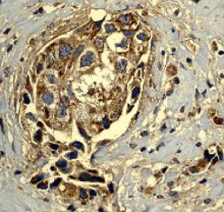
Anti-Hsp60 antibody
| Name | Anti-Hsp60 antibody |
|---|---|
| Supplier | Abcam |
| Catalog | ab46798 |
| Prices | $403.00 |
| Sizes | 100 µl |
| Host | Rabbit |
| Clonality | Polyclonal |
| Isotype | IgG |
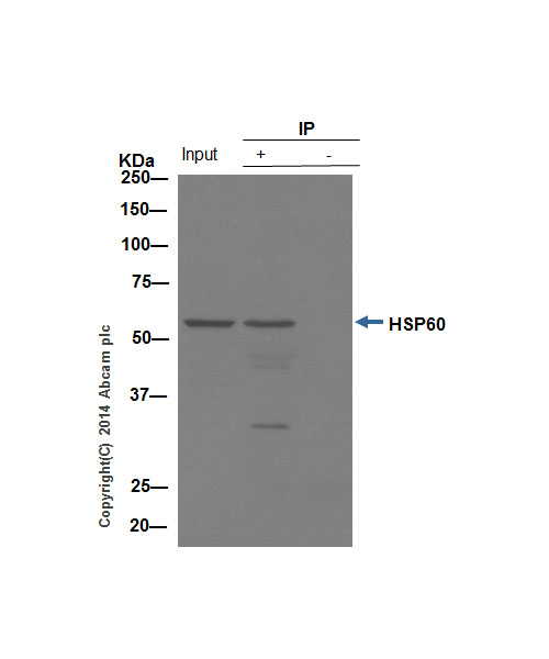
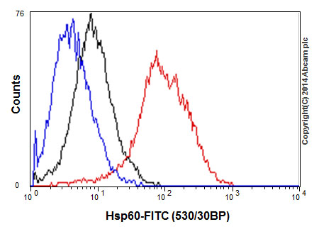
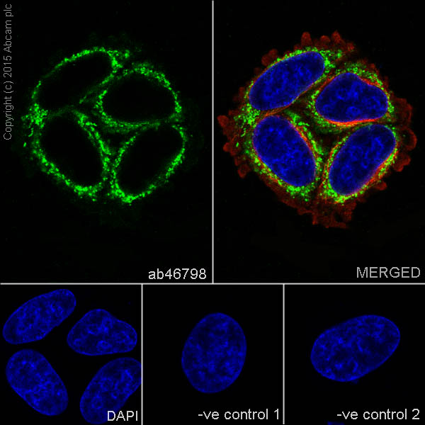
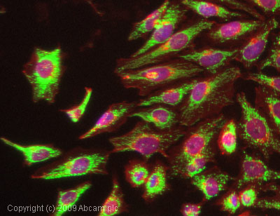
| Applications | IHC-F WB IHC-P ICC/IF ICC/IF FC IP |
| Species Reactivities | Mouse, Rat, Hamster, Human, Pig, Chicken, Bovine, Arabidopsis thaliana, Chlamydomonas reinhardtii, Orangutan |
| Antigen | Synthetic peptide (the amino acid sequence is considered to be commercially sensitive) corresponding to Human Hsp60 aa 400-500 |
| Description | Rabbit Polyclonal |
| Gene | HSPD1 |
| Conjugate | Unconjugated |
| Supplier Page | Shop |
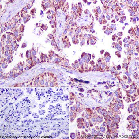
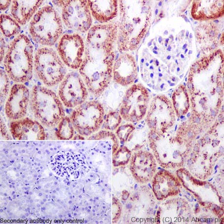
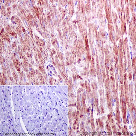
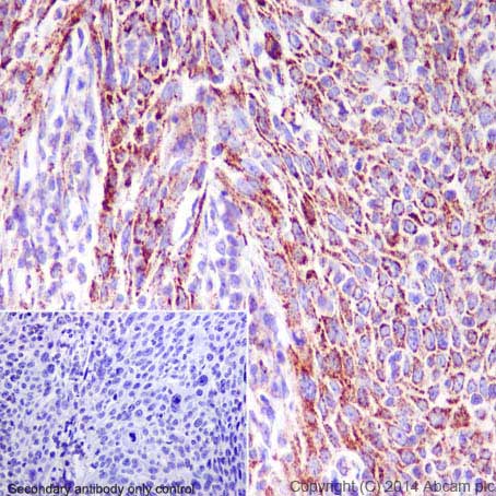
Product images
Product References
Active hexose-correlated compound down-regulates HSP27 of pancreatic cancer - Active hexose-correlated compound down-regulates HSP27 of pancreatic cancer
Suenaga S, Kuramitsu Y, Kaino S, Maehara S, Maehara Y, Sakaida I, Nakamura K. Anticancer Res. 2014 Jan;34(1):141-6.
Post-translational decrease in respiratory chain proteins in the Polg mutator - Post-translational decrease in respiratory chain proteins in the Polg mutator
Hauser DN, Dillman AA, Ding J, Li Y, Cookson MR. PLoS One. 2014 Apr 10;9(4):e94646.
Mitophagy enhances oncolytic measles virus replication by mitigating - Mitophagy enhances oncolytic measles virus replication by mitigating
Xia M, Gonzalez P, Li C, Meng G, Jiang A, Wang H, Gao Q, Debatin KM, Beltinger C, Wei J. J Virol. 2014 May;88(9):5152-64.
The mitochondrial protein NLRX1 controls the balance between extrinsic and - The mitochondrial protein NLRX1 controls the balance between extrinsic and
Soares F, Tattoli I, Rahman MA, Robertson SJ, Belcheva A, Liu D, Streutker C, Winer S, Winer DA, Martin A, Philpott DJ, Arnoult D, Girardin SE. J Biol Chem. 2014 Jul 11;289(28):19317-30.
Proteomic analysis of lipid droplets from Caco-2/TC7 enterocytes identifies novel - Proteomic analysis of lipid droplets from Caco-2/TC7 enterocytes identifies novel
Beilstein F, Bouchoux J, Rousset M, Demignot S. PLoS One. 2013;8(1):e53017.
.
Placental and vascular adaptations to exercise training before and during - Placental and vascular adaptations to exercise training before and during
Gilbert JS, Banek CT, Bauer AJ, Gingery A, Dreyer HC. Am J Physiol Regul Integr Comp Physiol. 2012 Sep 1;303(5):R520-6. doi:
Mitochondrial nucleoid interacting proteins support mitochondrial protein - Mitochondrial nucleoid interacting proteins support mitochondrial protein
He J, Cooper HM, Reyes A, Di Re M, Sembongi H, Litwin TR, Gao J, Neuman KC, Fearnley IM, Spinazzola A, Walker JE, Holt IJ. Nucleic Acids Res. 2012 Jul;40(13):6109-21.
OPA1 links human mitochondrial genome maintenance to mtDNA replication and - OPA1 links human mitochondrial genome maintenance to mtDNA replication and
Elachouri G, Vidoni S, Zanna C, Pattyn A, Boukhaddaoui H, Gaget K, Yu-Wai-Man P, Gasparre G, Sarzi E, Delettre C, Olichon A, Loiseau D, Reynier P, Chinnery PF, Rotig A, Carelli V, Hamel CP, Rugolo M, Lenaers G. Genome Res. 2011 Jan;21(1):12-20.
Actin and myosin contribute to mammalian mitochondrial DNA maintenance. - Actin and myosin contribute to mammalian mitochondrial DNA maintenance.
Reyes A, He J, Mao CC, Bailey LJ, Di Re M, Sembongi H, Kazak L, Dzionek K, Holmes JB, Cluett TJ, Harbour ME, Fearnley IM, Crouch RJ, Conti MA, Adelstein RS, Walker JE, Holt IJ. Nucleic Acids Res. 2011 Jul;39(12):5098-108.
