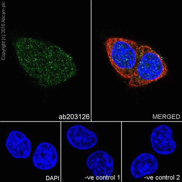
Immunofluorescent analysis of 4% paraformaldehyde-fixed, 0.1% Triton X-100 permeabilized HeLa (Human epithelial cells from cervix adenocarcinoma) cells labeling Hsp90 alpha + beta with ab203126 at 1/200 dilution, followed by Goat anti-rabbit IgG (Alexa Fluor® 488) (ab150077) secondary antibody at 1/1000 dilution (green). Confocal image showing cytoplasm and nuclear staining on HeLa cell line. The nuclear counter stain is DAPI (blue). Tubulin is detected with ab7291 (anti-Tubulin mouse mAb) at 1/1000 dilution and ab150120 (AlexaFluor®594 Goat anti-Mouse secondary) at 1/1000 dilution (red).The negative controls are as follows:-ve control 1: ab203126 at 1/200 dilution followed by ab150120 (AlexaFluor®594 Goat anti-Mouse secondary) at 1/1000 dilution.-ve control 2: ab7291 (anti-Tubulin mouse mAb) at 1/1000 dilution followed by ab150077 (Alexa Fluor®488 Goat Anti-Rabbit IgG H&L) at 1/1000 dilution.
![All lanes : Anti-Hsp90 alpha + beta antibody [EPR16621-67] (ab203126) at 1/10000 dilutionLane 1 : Hsp90 alpha recombinant protein fragment (GST-tag): aa533-732Lane 2 : Hsp90 beta recombinant protein fragment (His-Tag®): aa525-724Lysates/proteins at 0.01 µg per lane.SecondaryGoat Anti-Rabbit IgG H&L (HRP) (ab97051) at 1/1000 dilution](http://www.bioprodhub.com/system/product_images/ab_products/2/sub_3/5161_ab203126-245255-wb-1.jpg)
All lanes : Anti-Hsp90 alpha + beta antibody [EPR16621-67] (ab203126) at 1/10000 dilutionLane 1 : Hsp90 alpha recombinant protein fragment (GST-tag): aa533-732Lane 2 : Hsp90 beta recombinant protein fragment (His-Tag®): aa525-724Lysates/proteins at 0.01 µg per lane.SecondaryGoat Anti-Rabbit IgG H&L (HRP) (ab97051) at 1/1000 dilution
![All lanes : Anti-Hsp90 alpha + beta antibody [EPR16621-67] (ab203126) at 1/10000 dilutionLane 1 : Human fetal brain lysateLane 2 : Human fetal heart lysateLane 3 : Human fetal kidney lysateLysates/proteins at 10 µg per lane.SecondaryAnti-Rabbit IgG (HRP), specific to the non-reduced form of IgG at 1/1000 dilution](http://www.bioprodhub.com/system/product_images/ab_products/2/sub_3/5162_ab203126-245254-wb-2.jpg)
All lanes : Anti-Hsp90 alpha + beta antibody [EPR16621-67] (ab203126) at 1/10000 dilutionLane 1 : Human fetal brain lysateLane 2 : Human fetal heart lysateLane 3 : Human fetal kidney lysateLysates/proteins at 10 µg per lane.SecondaryAnti-Rabbit IgG (HRP), specific to the non-reduced form of IgG at 1/1000 dilution
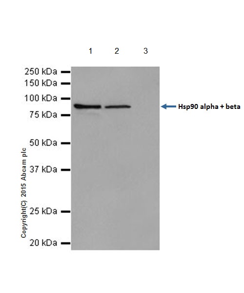
![Lanes 1, 3 & 4 : Anti-Hsp90 alpha + beta antibody [EPR16621-67] (ab203126) at 1/100000 dilutionLane 2 : Anti-Hsp90 alpha + beta antibody [EPR16621-67] (ab203126) at 1/10000 dilutionLane 1 : HeLa (Human epithelial cells from cervix adenocarcinoma) whole cell lysateLane 2 : HEK-293 (Human epithelial cells from embryonic kidney) whole cell lysateLane 3 : K562 (Human chronic myelogenous leukemia cells from bone marrow) whole cell lysateLane 4 : Jurkat (Human T cell leukemia cells from peripheral blood) whole cell lysateLysates/proteins at 20 µg per lane.SecondaryGoat Anti-Rabbit IgG H&L (HRP) (ab97051) at 1/1000 dilution](http://www.bioprodhub.com/system/product_images/ab_products/2/sub_3/5163_ab203126-245253-wb-3.jpg)
Lanes 1, 3 & 4 : Anti-Hsp90 alpha + beta antibody [EPR16621-67] (ab203126) at 1/100000 dilutionLane 2 : Anti-Hsp90 alpha + beta antibody [EPR16621-67] (ab203126) at 1/10000 dilutionLane 1 : HeLa (Human epithelial cells from cervix adenocarcinoma) whole cell lysateLane 2 : HEK-293 (Human epithelial cells from embryonic kidney) whole cell lysateLane 3 : K562 (Human chronic myelogenous leukemia cells from bone marrow) whole cell lysateLane 4 : Jurkat (Human T cell leukemia cells from peripheral blood) whole cell lysateLysates/proteins at 20 µg per lane.SecondaryGoat Anti-Rabbit IgG H&L (HRP) (ab97051) at 1/1000 dilution
![All lanes : Anti-Hsp90 alpha + beta antibody [EPR16621-67] (ab203126) at 1/10000 dilutionLane 1 : Mouse brain lysateLane 2 : Mouse heart lysateLane 3 : Mouse kidney lysateLane 4 : Rat brain lysateLane 5 : Rat heart lysateLane 6 : Rat kidney lysateLane 7 : C6 (Rat glial tumor cells) whole cell lysateLane 8 : RAW 264.7 (Mouse macrophage cells transformed with Abelson murine leukemia virus) whole cell lysateLane 9 : PC-12 (Rat adrenal gland pheochromocytoma) whole cell lysateLane 10 : NIH/3T3 (Mouse embyro fibroblast cells) whole cell lysateLysates/proteins at 10 µg per lane.SecondaryAnti-Rabbit IgG (HRP), specific to the non-reduced form of IgG at 1/1000 dilution](http://www.bioprodhub.com/system/product_images/ab_products/2/sub_3/5164_ab203126-245252-wb-4.jpg)
All lanes : Anti-Hsp90 alpha + beta antibody [EPR16621-67] (ab203126) at 1/10000 dilutionLane 1 : Mouse brain lysateLane 2 : Mouse heart lysateLane 3 : Mouse kidney lysateLane 4 : Rat brain lysateLane 5 : Rat heart lysateLane 6 : Rat kidney lysateLane 7 : C6 (Rat glial tumor cells) whole cell lysateLane 8 : RAW 264.7 (Mouse macrophage cells transformed with Abelson murine leukemia virus) whole cell lysateLane 9 : PC-12 (Rat adrenal gland pheochromocytoma) whole cell lysateLane 10 : NIH/3T3 (Mouse embyro fibroblast cells) whole cell lysateLysates/proteins at 10 µg per lane.SecondaryAnti-Rabbit IgG (HRP), specific to the non-reduced form of IgG at 1/1000 dilution
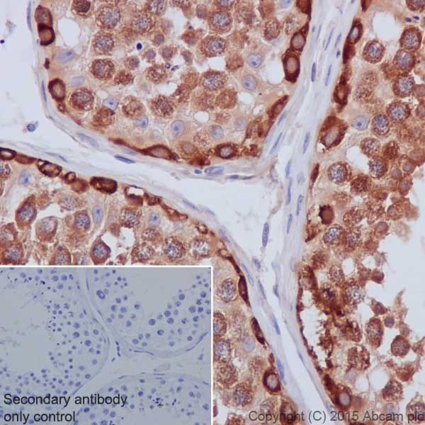
Immunohistochemical analysis of paraffin-embedded Human testis tissue labeling Hsp90 alpha + beta with ab203126 at 1/1000 dilution, followed by Goat Anti-Rabbit IgG H&L (HRP) (ab97051) secondary antibody at 1/500 dilution. Cytoplasm and weak nucleus staining on germ cells of Human testis is observed. Counter stained with Hematoxylin.Secondary antibody only control: Used PBS instead of primary antibody, secondary antibody is Goat Anti-Rabbit IgG H&L (HRP) (ab97051) at 1/500 dilution.
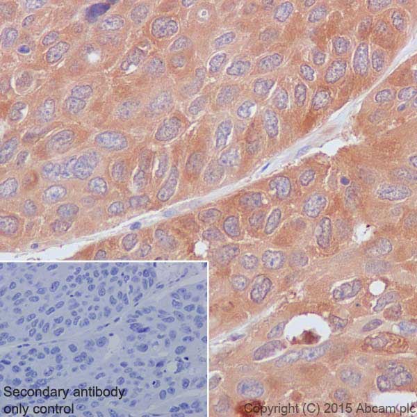
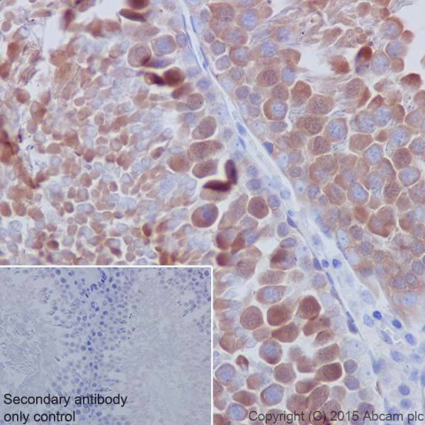
Immunohistochemical analysis of paraffin-embedded Human lung cancer tissue labeling Hsp90 alpha + beta with ab203126 at 1/1000 dilution, followed by Goat Anti-Rabbit IgG H&L (HRP) (ab97051) secondary antibody at 1/500 dilution. Cytoplasm and weak nucleus staining on tumor cells of Human lung cancer is observed. Counter stained with Hematoxylin.Secondary antibody only control: Used PBS instead of primary antibody, secondary antibody is Goat Anti-Rabbit IgG H&L (HRP) (ab97051) at 1/500 dilution.
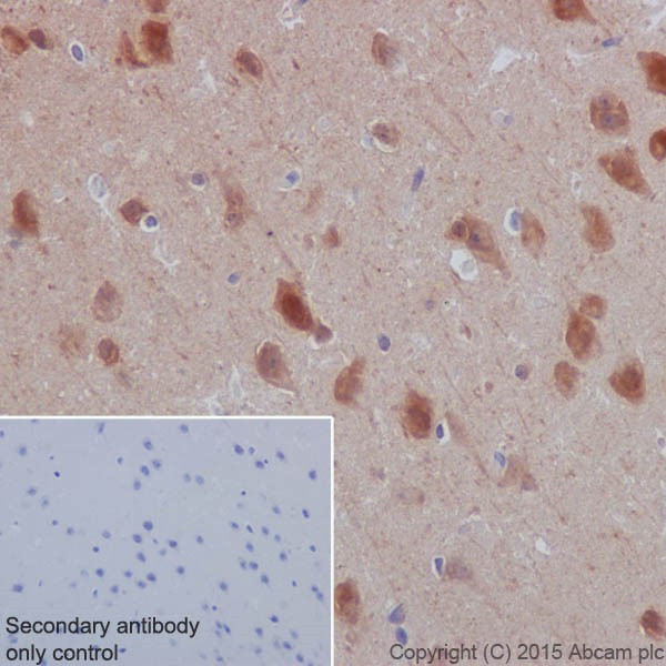
Immunohistochemical analysis of paraffin-embedded Mouse cerebral cortex tissue labeling Hsp90 alpha + beta with ab203126 at 1/1000 dilution, followed by Goat Anti-Rabbit IgG H&L (HRP) (ab97051) secondary antibody at 1/500 dilution. Cytoplasm and nucleus staining on neuron of mouse cerebral cortex is observed. Counter stained with Hematoxylin.Secondary antibody only control: Used PBS instead of primary antibody, secondary antibody is Goat Anti-Rabbit IgG H&L (HRP) (ab97051) at 1/500 dilution.
Immunohistochemical analysis of paraffin-embedded rat testis tissue labeling Hsp90 alpha + beta with ab203126 at 1/1000 dilution, followed by Goat Anti-Rabbit IgG H&L (HRP) (ab97051) secondary antibody at 1/500 dilution. Cytoplasm and weak nucleus staining on germ cells of rat testis is observed. Counter stained with Hematoxylin.Secondary antibody only control: Used PBS instead of primary antibody, secondary antibody is Goat Anti-Rabbit IgG H&L (HRP) (ab97051) at 1/500 dilution.
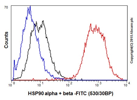
Flow cytometric analysis of 2% paraformaldehyde-fixed Jurkat (Human T cell leukemia cells from peripheral blood) cells labeling Hsp90 alpha + beta with ab203126 at 1/350 dilution (red) compared with a rabbit monoclonal IgG isotype control (ab172730; black) and an unlabelled control (cells without incubation with primary antibody and secondary antibody; blue). Goat anti rabbit IgG (FITC) at 1/150 dilution was used as the secondary antibody.
Hsp90 alpha + beta was immunoprecipitated from 1mg of HeLa (Human epithelial cells from cervix adenocarcinoma) whole cell lysate with ab203126 at 1/100 dilution. Western blot was performed from the immunoprecipitate using ab203126 at 1/1000 dilution. Anti-Rabbit IgG (HRP), specific to the non-reduced form of IgG, was used as secondary antibody at 1/1500 dilution.Lane 1: HeLa whole cell lysate 10 µg (Input).Lane 2: ab203126 IP in HeLa whole cell lysate.Lane 3: Rabbit monoclonal IgG (ab172730) instead of ab203126 in HeLa whole cell lysate.Blocking and dilution buffer and concentration: 5% NFDM/TBST.Exposure time: 3 seconds.