Anti-IL6 antibody (ab83339) at 1 µg/ml + Spleen (Mouse) Tissue Lysate at 10 µgSecondaryGoat polyclonal to Rabbit IgG - H&L - Pre-Adsorbed (HRP) at 1/3000 dilutiondeveloped using the ECL techniquePerformed under reducing conditions.

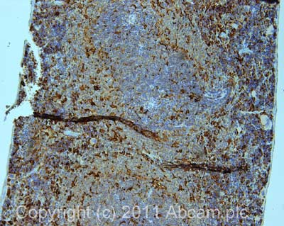
IHC image of IL6 staining in mouse spleen formalin fixed paraffin embedded tissue section, performed on a Leica BondTM system using the standard protocol F. The section was pre-treated using heat mediated antigen retrieval with sodium citrate buffer (pH6, epitope retrieval solution 1) for 20 mins. The section was then incubated with ab83339, 1µg/ml, for 15 mins at room temperature and detected using an HRP conjugated compact polymer system. DAB was used as the chromogen. The section was then counterstained with haematoxylin and mounted with DPX.

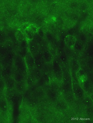
IHC-FoFr image of IL6 staining on rat hippocampal sections using ab83339 (1/300). This staining is consistent with observation made by the article reference 7805281. The sections used came from animals perfused fixed with Paraformaldehyde 4% with 15% of a solution of saturated picric acid, in phosphate buffer 0.1M. Following postfixation in the same fixative overnight, the spinal cord were cryoprotected in sucrose 30% overnight. Spinal cords were then cut using a cryostat and the immunostainings were performed using the ‘free floating’ technique.See Abreview


