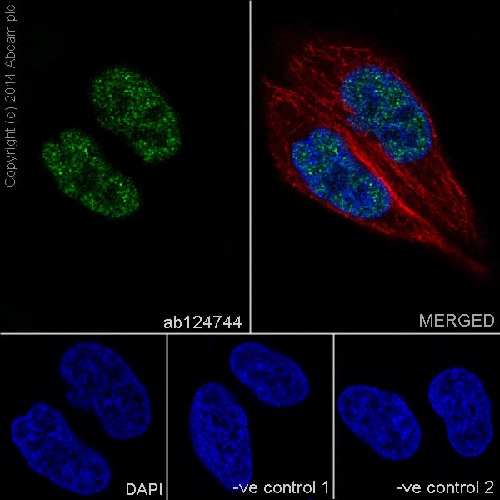
Immunofluorescent analysis of 4% paraformaldehyde-fixed, 0.1% Triton X-100 permeabilized HeLa (Human epithelial cells from cervix adenocarcinoma) cells labeling IRF2 with ab124744 at 1/500 dilution, followed by Goat anti-rabbit IgG (Alexa Fluor® 488) (ab150077) secondary antibody at 1/500 dilution (green). Confocal image showing nuclear staining on HeLa cells.The nuclear counter stain is DAPI (blue). Tubulin is detected with ab7291 (anti-Tubulin mouse mAb) at 1/1000 dilution and ab150120 (AlexaFluor®594 Goat anti-Mouse secondary) at 1/500 dilution (red).The negative controls are as follows:-ve control 1: ab124744 at 1/500 dilution followed by ab150120 (AlexaFluor®594 Goat anti-Mouse secondary) at 1/500 dilution.-ve control 2: ab7291 (anti-Tubulin mouse mAb) at 1/1000 dilution followed by ab150077 (Alexa Fluor®488 Goat Anti-Rabbit IgG H&L) at 1/500 dilution.
![All lanes : Anti-IRF2 antibody [EPR4644(2)] (ab124744) at 1/5000 dilutionLane 1 : SW480 (Human colorectal adenocarcinoma cell line)Lane 2 : HeLa (Human epithelial cells from cervix adenocarcinoma)Lane 3 : Jurkat (Human T cell leukemia cells from peripheral blood)Lysates/proteins at 20 µg per lane.SecondaryGoat Anti-Rabbit IgG, (H+L),Peroxidase conjugated at 1/1000 dilution](http://www.bioprodhub.com/system/product_images/ab_products/2/sub_3/9971_ab124744-242983-ab124744.jpg)
All lanes : Anti-IRF2 antibody [EPR4644(2)] (ab124744) at 1/5000 dilutionLane 1 : SW480 (Human colorectal adenocarcinoma cell line)Lane 2 : HeLa (Human epithelial cells from cervix adenocarcinoma)Lane 3 : Jurkat (Human T cell leukemia cells from peripheral blood)Lysates/proteins at 20 µg per lane.SecondaryGoat Anti-Rabbit IgG, (H+L),Peroxidase conjugated at 1/1000 dilution
![All lanes : Anti-IRF2 antibody [EPR4644(2)] (ab124744) at 1/1000 dilutionLane 1 : Caco-2 (Human colorectal adenocarcinoma cells)Lane 2 : RAW 264.7 (Mouse macrophage cells transformed with Abelson murine leukemia virus)Lane 3 : NIH/3T3 (Mouse embyro fibroblast cells)Lysates/proteins at 10 µg per lane.SecondaryGoat Anti-Rabbit IgG, (H+L),Peroxidase conjugated at 1/1000 dilution](http://www.bioprodhub.com/system/product_images/ab_products/2/sub_3/9972_ab124744-242900-ab124744.jpg)
All lanes : Anti-IRF2 antibody [EPR4644(2)] (ab124744) at 1/1000 dilutionLane 1 : Caco-2 (Human colorectal adenocarcinoma cells)Lane 2 : RAW 264.7 (Mouse macrophage cells transformed with Abelson murine leukemia virus)Lane 3 : NIH/3T3 (Mouse embyro fibroblast cells)Lysates/proteins at 10 µg per lane.SecondaryGoat Anti-Rabbit IgG, (H+L),Peroxidase conjugated at 1/1000 dilution
![Anti-IRF2 antibody [EPR4644(2)] (ab124744) at 1/1000 dilution + Human fetal lung lysate at 10 µgSecondaryAnti-Rabbit IgG (HRP), specific to the non-reduced form of IgG at 1/1000 dilution](http://www.bioprodhub.com/system/product_images/ab_products/2/sub_3/9973_ab124744-242901-ab124744.jpg)
Anti-IRF2 antibody [EPR4644(2)] (ab124744) at 1/1000 dilution + Human fetal lung lysate at 10 µgSecondaryAnti-Rabbit IgG (HRP), specific to the non-reduced form of IgG at 1/1000 dilution
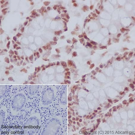
Immunohistochemistry (Formalin/PFA-fixed paraffin-embedded sections) analysis of Human colon tissue labeling IRF2 with purified ab124744 at 1/50. Heat mediated antigen retrieval was performed using Tris/EDTA buffer pH 9. Goat Anti-Rabbit IgG H&L (HRP) (ab97051) at 1/500 dilution was used as the secondary antibody. Nucleus staining on epithelium of human colon was observed. Negative control using PBS instead of primary antibody. Counterstained with Hematoxylin.
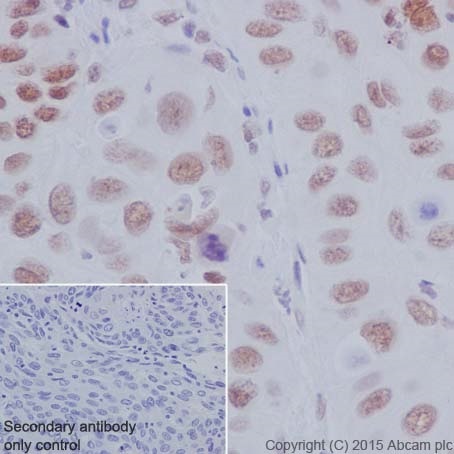
Immunohistochemistry (Formalin/PFA-fixed paraffin-embedded sections) analysis of Human cervical cancer tissue labeling IRF2 with purified ab124744 at 1/50. Heat mediated antigen retrieval was performed using Tris/EDTA buffer pH 9. Goat Anti-Rabbit IgG H&L (HRP) (ab97051) at 1/500 dilution was used as the secondary antibody. Nucleus staining on tumor cells of human cervix cancer was observed. Negative control using PBS instead of primary antibody. Counterstained with Hematoxylin.
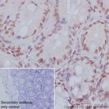
Immunohistochemistry (Formalin/PFA-fixed paraffin-embedded sections) analysis of Mouse colon tissue labeling IRF2 with purified ab124744 at 1/50. Heat mediated antigen retrieval was performed using Tris/EDTA buffer pH 9. Goat Anti-Rabbit IgG H&L (HRP) (ab97051) at 1/500 dilution was used as the secondary antibody. Nucleus staining on epithelial cells of mouse colon was observed. Negative control using PBS instead of primary antibody. Counterstained with Hematoxylin.
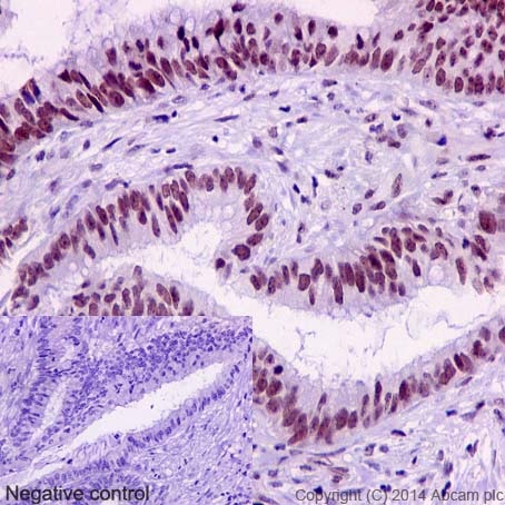
Immunohistochemistry (Formalin/PFA-fixed paraffin-embedded sections) analysis of human colonic carcinoma tissue labelling IRF2 with purified ab124744 at 1/50. Heat mediated antigen retrieval was performed using Tris/EDTA buffer pH 9. A prediluted HRP-polymer conjugated anti-rabbit IgG was used as the secondary antibody. Negative control using PBS instead of primary antibody. Counterstained with Hematoxylin.
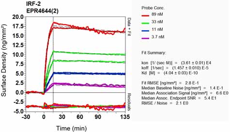
Equilibrium disassociation constant (KD)Learn more about KD Click here to learn more about KD

![All lanes : Anti-IRF2 antibody [EPR4644(2)] (ab124744) at 1/5000 dilutionLane 1 : SW480 (Human colorectal adenocarcinoma cell line)Lane 2 : HeLa (Human epithelial cells from cervix adenocarcinoma)Lane 3 : Jurkat (Human T cell leukemia cells from peripheral blood)Lysates/proteins at 20 µg per lane.SecondaryGoat Anti-Rabbit IgG, (H+L),Peroxidase conjugated at 1/1000 dilution](http://www.bioprodhub.com/system/product_images/ab_products/2/sub_3/9971_ab124744-242983-ab124744.jpg)
![All lanes : Anti-IRF2 antibody [EPR4644(2)] (ab124744) at 1/1000 dilutionLane 1 : Caco-2 (Human colorectal adenocarcinoma cells)Lane 2 : RAW 264.7 (Mouse macrophage cells transformed with Abelson murine leukemia virus)Lane 3 : NIH/3T3 (Mouse embyro fibroblast cells)Lysates/proteins at 10 µg per lane.SecondaryGoat Anti-Rabbit IgG, (H+L),Peroxidase conjugated at 1/1000 dilution](http://www.bioprodhub.com/system/product_images/ab_products/2/sub_3/9972_ab124744-242900-ab124744.jpg)
![Anti-IRF2 antibody [EPR4644(2)] (ab124744) at 1/1000 dilution + Human fetal lung lysate at 10 µgSecondaryAnti-Rabbit IgG (HRP), specific to the non-reduced form of IgG at 1/1000 dilution](http://www.bioprodhub.com/system/product_images/ab_products/2/sub_3/9973_ab124744-242901-ab124744.jpg)


