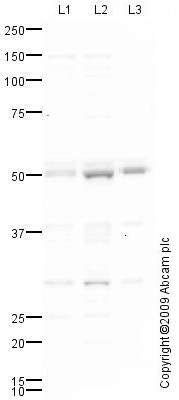
All lanes : Anti-IRF3 antibody (ab76367) at 1 µg/mlLane 1 : HeLa (Human epithelial carcinoma cell line) Whole Cell Lysate Lane 2 : Jurkat (Human T cell lymphoblast-like cell line) Whole Cell LysateLane 3 : THP1 (Human acute monocytic leukemia cell line) Whole Cell Lysate Lysates/proteins at 10 µg per lane.SecondaryGoat polyclonal to Rabbit IgG - H&L - Pre-Adsorbed (HRP) at 1/3000 dilutiondeveloped using the ECL techniquePerformed under reducing conditions.
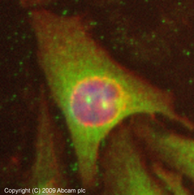
ICC/IF image of ab76367 stained HeLa cells. The cells were 4% PFA fixed (10 min) and then incubated in 1%BSA / 10% normal goat serum / 0.3M glycine in 0.1% PBS-Tween for 1h to permeabilise the cells and block non-specific protein-protein interactions. The cells were then incubated with the antibody (ab76367, 5µg/ml) overnight at +4°C. The secondary antibody (green) was Alexa Fluor® 488 goat anti-rabbit IgG (H+L) used at a 1/1000 dilution for 1h. Alexa Fluor® 594 WGA was used to label plasma membranes (red) at a 1/200 dilution for 1h. DAPI was used to stain the cell nuclei (blue) at a concentration of 1.43µM. This antibody also gave a positive result in 4% PFA fixed (10 min) MCF7 cells at 5µg/ml, and in 100% methanol fixed (5 min) HeLa, Hek293, HepG2 and MCF7 cells at 5µg/ml.
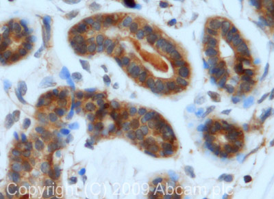
IHC image of IRF3 staining in human breast carcinoma formalin fixed paraffin embedded tissue section, performed on a Leica BondTM system using the standard protocol F. The section was pre-treated using heat mediated antigen retrieval with sodium citrate buffer (pH6, epitope retrieval solution 1) for 20 mins. The section was then incubated with ab76367, 5µg/ml, for 15 mins at room temperature and detected using an HRP conjugated compact polymer system. DAB was used as the chromogen. The section was then counterstained with haematoxylin and mounted with DPX.