E-Cadherin (24E10) Rabbit mAb
| Name | E-Cadherin (24E10) Rabbit mAb |
|---|---|
| Supplier | Cell Signaling Technology |
| Catalog | 3195 |
| Prices | $99.00, $246.00 |
| Sizes | 20 µl (2 western blots), 100 µl (10 western blots) |
| Host | Rabbit |
| Clonality | Monoclonal |
| Isotype | IgG |
| Clone | 24E10 |
| Applications | WB IHC-P IHC-F ICC/IF FC |
| Species Reactivities | Human, Mouse, Bovine, Dog, Pig |
| Antigen | Monoclonal antibody is produced by immunizing animals with a synthetic peptide corresponding to the sequence surrounding Pro780 of human E-cadherin protein. |
| Description | Rabbit Monoclonal |
| Gene | CDH1 |
| Supplier Page | Shop |
Product images
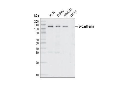 Western blot analysis of extracts from various cell lines, using E-Cadherin (24E10) Rabbit mAb.
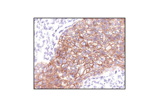
 Immunohistochemical analysis of paraffin-embedded human lung carcinoma, using E-Cadherin (24E10) Rabbit mAb.
 Immunohistochemical analysis of paraffin-embedded human metastatic adenocarcinoma in lymph node, using E-Cadherin (24E10) Rabbit mAb.
 Immunohistochemical analysis of paraffin-embedded mouse lung using E-Cadherin (24E10) Rabbit mAb.
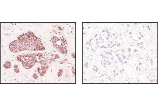 Immunohistochemical analysis of paraffin-embedded human breast carcinoma, using E-Cadherin (24E10) Rabbit mAb in the presence of control peptide (left) or E-Cadherin Blocking Peptide #1050 (right).
 Immunohistochemical analysis of frozen HCC827 xenograft, showing membrane and cytoplasmic localization using E-Cadherin (24E10) Rabbit mAb.
 Confocal immunofluorescent images of MCF7 cells using E-Cadherin (24E10) Rabbit mAb (green, left) compared to an isotype control (right). Blue pseudocolor = DRAQ5 ® (fluorescent DNA dye).
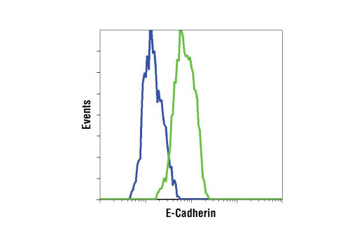 Flow cytometric analysis of HeLa cells (blue) and MCF7 cells (green) using E-Cadherin (24E10) Rabbit mAb.