
All lanes : Anti-Ki67 antibody (ab15580) at 1 µg/mlLane 1 : Hela whole cell lysateLane 2 : Hela whole cell lysate with Ki67 peptide (ab15581) at 1 µg/mlLysates/proteins at 20 µg per lane.SecondaryAlexa Fluor Goat polyclonal to Rabbit IgG at 1/10000 dilutionPerformed under reducing conditions.

Fluorescent confocal microscopy (20x) of mouse (P0) olfactory bulb, outer glomeruli layer, showing Ki67 immunoreactivity (ab15580; 1/1000; overnight at RT, 0.25% TX-100 no blocking step) using a secondary goat anti-rabbit fluorescent antibody (Alexa Fluor 488;1/300 2h at RT.
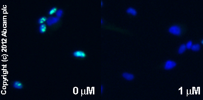
ab15580 staining Ki67 in SK-N-SH cells treated with NADA (N-Arachidonyldopamine) (ab120099), by ICC/IF. Decrease in Ki67 expression correlates with increased concentration of NADA (N-Arachidonyldopamine), as described in literature.The cells were incubated at 37°C for 10 minutes in media containing different concentrations of ab120099 (NADA (N-Arachidonyldopamine)) in ethanol, fixed with 4% formaldehyde for 10 minutes at room temperature and blocked with PBS containing 10% goat serum, 0.3 M glycine, 1% BSA and 0.1% tween for 2h at room temperature. Staining of the treated cells with ab15580 (1 µg/ml) was performed overnight at 4°C in PBS containing 1% BSA and 0.1% tween. A DyLight 488 goat anti-rabbit polyclonal antibody (ab96899) at 1/250 dilution was used as the secondary antibody. Nuclei were counterstained with DAPI and are shown in blue.

SK-N-SH cells were permitted to grow to confluency, then serum starved for 48 hours and predominantly driven into G0. The cells were then paraformaldehyde fixed and immunofluorescently labelled with anti-Ki67 (ab15580) at a dilution of 1/1000. The majority of the cells show little or no Ki67 staining, indicating they are in G0 arrest (red cells). Two cells however show strong nucleolar Ki67 staining indicating they are still cycling (green cells). The DNA is stained with DAPI and is shown in red. The Ki67 staining is shown in green. x 63 magnification.Similar results were seen with an asynchronous population of HeLa cells. The Ki67 staining was localised to the periphery of the nucleoli and throughout the nucleoplasm of proliferating cells. (This data is not shown but is available upon request).

Immunostaining for Rabbit polyclonal to Ki67 - Proliferation Marker (ab15580) on Mouse P7 brain (frozen) sections. Sections were fixed with paraformaldehyde, neither a blocking nor antigen retrieval steps were included in this protocol. Secondary antibody used was Rabbit IgG antibody (ab7082; 1/250).

ab15580 at 1/50 staining Human umbilical artery endothelial cells by ICC/IF. The tissue was paraformaldehyde fixed and blocked before permeabilization with saponin and incubation with the antibody for 16 hours. A FITC conjugated goat anti-rabbit IgG was used as the secondary. The merged image shows those cells expressing Ki67 from the total number of exponential cells.See Abreview
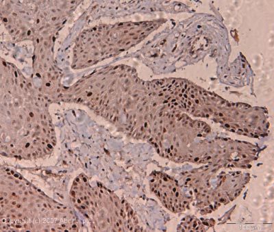
IHC image of ab15580 stained human skin carcinoma FFPE section. Section was pre-treated using pressure cooker heat mediated antigen retrieval with sodium citrate buffer (pH6) for 30 seconds at 125°C. Section was incubated with ab15580 at a dilution of 1:200 for 1h at room temperature and detected using an HRP conjugated polymer system. DAB was used as the chromogen. The section was then counterstained with haematoxylin and mounted with DPX.
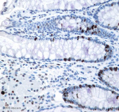
ab15580 staining Ki67-Proliferation Maker in human colon tissue sections by IHC-P (formaldehyde-fixed paraffin-embedded sections). Tissue samples were fixed with formaldehyde and blocked with 10% goat serum for 30 minutes at 25°C. Antigen retrieval was by heat mediation in Target Retrieval Solution. Samples were incubated with primary antibody 1/5000 (TBST) for 1 hour at 25°C. See Abreview
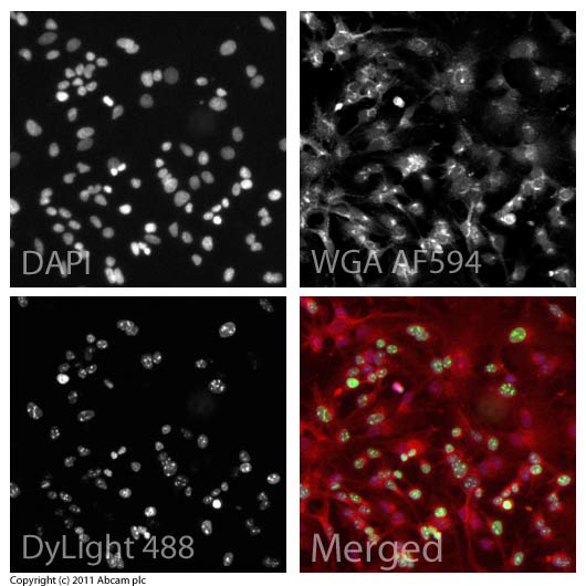
ICC/IF image of ab15580 stained HepG2 cells. The cells were 100% methanol fixed (5 min) and then incubated in 1%BSA / 10% normal goat serum / 0.3M glycine in 0.1% PBS-Tween for 1h to permeabilise the cells and block non-specific protein-protein interactions. The cells were then incubated with the antibody (ab15580, 1µg/ml) overnight at +4°C. The secondary antibody (green) was ab96899, DyLight® 488 goat anti-rabbit IgG (H+L) used at a 1/250 dilution for 1h. Alexa Fluor® 594 WGA was used to label plasma membranes (red) at a 1/200 dilution for 1h. DAPI was used to stain the cell nuclei (blue) at a concentration of 1.43µM.
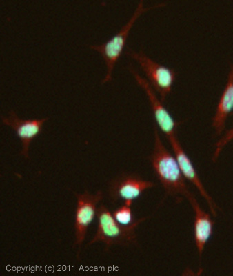
ICC/IF image of ab15580 stained MEF1 cells. The cells were 4% formaldehyde fixed (10 min) and then incubated in 1%BSA / 10% normal goat serum / 0.3M glycine in 0.1% PBS-Tween for 1h to permeabilise the cells and block non-specific protein-protein interactions. The cells were then incubated with the antibody (ab15580, 1µg/ml) overnight at +4°C. The secondary antibody (green) was ab96899, DyLight® 488 goat anti-rabbit IgG (H+L) used at a 1/250 dilution for 1h. Alexa Fluor® 594 WGA was used to label plasma membranes (red) at a 1/200 dilution for 1h. DAPI was used to stain the cell nuclei (blue) at a concentration of 1.43µM.
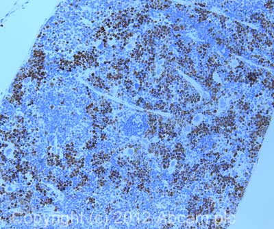
IHC image of ab15580 staining in mouse spleen formalin fixed paraffin embedded tissue section, performed on a Leica BondTM system using the standard protocol B. The section was pre-treated using heat mediated antigen retrieval with sodium citrate buffer (pH6, epitope retrieval solution 1) for 20 mins. The section was then incubated with ab15580, 1µg/ml, for 15 mins at room temperature. A goat anti-rabbit biotinylated secondary antibody was used to detect the primary, and visualized using an HRP conjugated ABC system. DAB was used as the chromogen. The section was then counterstained with haematoxylin and mounted with DPX. For other IHC staining systems (automated and non-automated) customers should optimize variable parameters such as antigen retrieval conditions, primary antibody concentration and antibody incubation times.
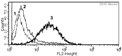
ab15580 staining Ki67 - Proliferation Marker in Human PBMCs by Flow Cytometry. Cells were isolated by Ficoll density separation, fixed in paraformaldehyde and permeabilized in 0.1% Saponin + PBS. The sample was incubated with the primary antibody (1/100 in PBS + 1% FCS and 0.09% sodium azide) for 1 hour at 4°C. A phycoerythrin-conjugated Goat anti-rabbit IgG (1/100) was used as the secondary antibody.Gating Strategy: Lymphocytes and Monocytes 1 = isotype; 2 = PBMCs untreated; 3 = PBMCs treated with PHA.See Abreview
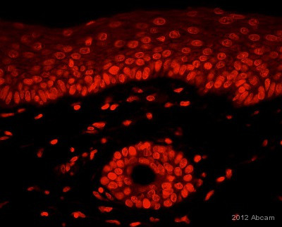
ab15580 staining Ki67 - Proliferation Marker in Human skin tissue sections by Immunohistochemistry (IHC-P - paraformaldehyde-fixed, paraffin-embedded sections). Tissue was fixed with paraformaldehyde and blocked with 4% serum for 30 minutes at 25°C; antigen retrieval was by heat mediation in a citrate buffer (pH 6.0). Samples were incubated with primary antibody (5 µg/ml in blocking buffer) for 16 hours at 4°C. A Texas Red ® Goat anti-rabbit IgG polyclonal (1/100) was used as the secondary antibody.See Abreview


