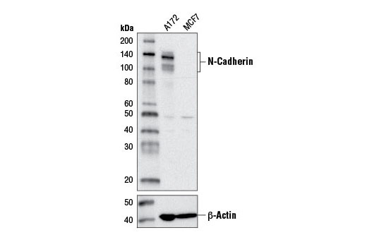
Western blot analysis of extracts from A172 and MCF7 cells using N-Cadherin (D4R1H) XP ® Rabbit mAb (upper) or β-Actin (D6A8) Rabbit mAb #8457 (lower).
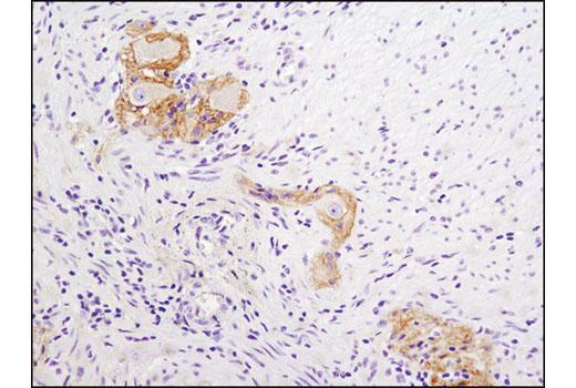
Immunohistochemical analysis of paraffin-embedded human colon using N-Cadherin (D4R1H) XP ® Rabbit mAb. Note staining of myenteric plexus.
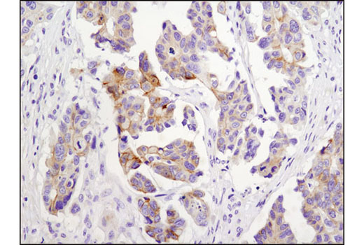
Immunohistochemical analysis of paraffin-embedded human ovarian carcinoma using N-Cadherin (D4R1H) XP ® Rabbit mAb.
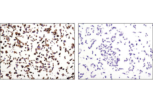
Immunohistochemical analysis of paraffin-embedded A172 (positive, left) and MCF7 (negative, right) cell pellets using N-Cadherin (D4R1H) XP ® Rabbit mAb.
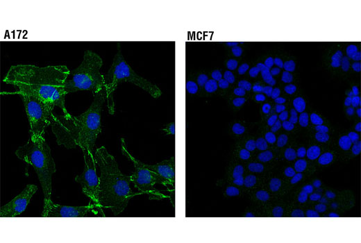
Confocal immunofluorescent analysis of A172 (positive, left) and MCF7 (negative, right) cells using N-Cadherin (D4R1H) XP ® Rabbit mAb (green). Blue pseudocolor= DRAQ5 ® #4084 (fluorescent DNA dye).


