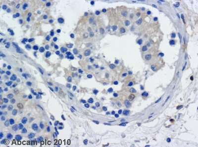
Anti-Kinesin Heavy Chain antibody [KN-01]
| Name | Anti-Kinesin Heavy Chain antibody [KN-01] |
|---|---|
| Supplier | Abcam |
| Catalog | ab9097 |
| Prices | $390.00 |
| Sizes | 100 µg |
| Host | Mouse |
| Clonality | Monoclonal |
| Isotype | IgM |
| Clone | KN-01 |
| Applications | IHC-P FC ICC/IF ICC/IF |
| Species Reactivities | Mouse, Human, Pig |
| Antigen | Enriched fraction of porcine brain kinesin |
| Description | Mouse Monoclonal |
| Gene | KIF5B |
| Conjugate | Unconjugated |
| Supplier Page | Shop |
Product images
Product References
Imaging type-III secretion reveals dynamics and spatial segregation of Salmonella - Imaging type-III secretion reveals dynamics and spatial segregation of Salmonella
Van Engelenburg SB, Palmer AE. Nat Methods. 2010 Apr;7(4):325-30.
KIF5B gene sequence variation and response of cardiac stroke volume to regular - KIF5B gene sequence variation and response of cardiac stroke volume to regular
Argyropoulos G, Stutz AM, Ilnytska O, Rice T, Teran-Garcia M, Rao DC, Bouchard C, Rankinen T. Physiol Genomics. 2009 Jan 8;36(2):79-88. doi:
Molecular motors implicated in the axonal transport of tau and alpha-synuclein. - Molecular motors implicated in the axonal transport of tau and alpha-synuclein.
Utton MA, Noble WJ, Hill JE, Anderton BH, Hanger DP. J Cell Sci. 2005 Oct 15;118(Pt 20):4645-54. Epub 2005 Sep 21.
Morphogenesis of the telencephalic commissure requires scaffold protein - Morphogenesis of the telencephalic commissure requires scaffold protein
Kelkar N, Delmotte MH, Weston CR, Barrett T, Sheppard BJ, Flavell RA, Davis RJ. Proc Natl Acad Sci U S A. 2003 Aug 19;100(17):9843-8. Epub 2003 Aug 1.
Monoclonal antibody KN-01 against the heavy chain of kinesin. - Monoclonal antibody KN-01 against the heavy chain of kinesin.
Draberova E, Macurek L, Richterova V, Bohm KJ, Draber P. Folia Biol (Praha). 2002;48(2):77-9.


![Overlay histogram showing HeLa cells stained with ab9097 (red line). The cells were fixed with 80% methanol (5 min) and then permeabilized with 0.1% PBS-Tween for 20 min. The cells were then incubated in 1x PBS / 10% normal goat serum / 0.3M glycine to block non-specific protein-protein interactions. The cells were then incubated with the antibody (ab90970, 1µg/1x106 cells) for 30 min at 22ºC. The secondary antibody used was DyLight® 488 goat anti-mouse IgG (H+L) (ab96879) at 1/500 dilution for 30 min at 22ºC. Isotype control antibody (black line) was mouse IgM [ICIGM] (ab91545, 2µg/1x106 cells ) used under the same conditions. Acquisition of >5,000 events was performed. This antibody gave a positive signal in HeLa cells fixed with 4% paraformaldehyde (10 min)/permeabilized in 0.1% PBS-Tween used under the same conditions.](http://www.bioprodhub.com/system/product_images/ab_products/2/sub_3/13132_Kinesin-Heavy-Chain-Primary-antibodies-ab9097-2.jpg)