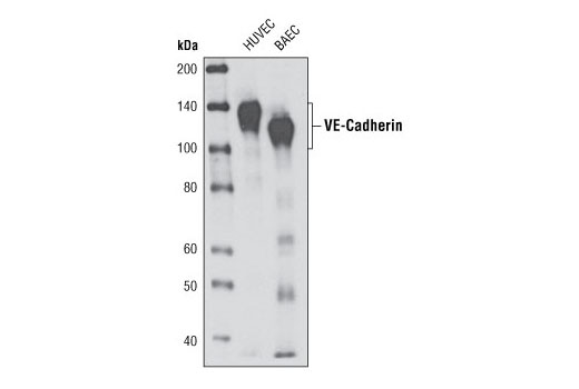
Western blot analysis of extracts from HUVEC and BAEC cells using VE-Cadherin (D87F2) XP ® Rabbit mAb.

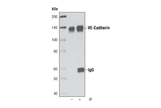
Immunoprecipitation of VE-caderin from HUVEC cells using VE-Cadherin (D87F2) XP ® Rabbit mAb followed by western blot using the same antibody. Lane 1 is 5% input.

Confocal immunofluorescent analysis of HUVE cells (left) and HeLa cells (right) using VE-Cadherin (D87F2) XP ® Rabbit mAb (green). Actin filaments have been labeled with DY-554 phalloidin (red). Blue pseudocolor = DRAQ5® #4084 (fluorescent DNA dye).

Flow cytometric analysis of HeLa cells (blue) and HUVEC cells (green) using VE-Cadherin (D87F2) Rabbit mAb.


