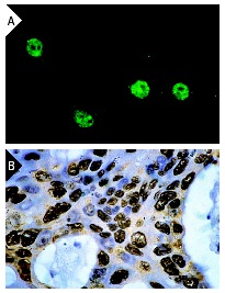
c-Jun (KM-1): sc-822. Immunofluorescence staining of methanol-fixed NIH/3T3 cells showing punctate nuclear staining of phosphorylated c-Jun (A). Immunoperoxidase staining of formalin-fixed, paraffin-embedded human colon carcinoma showing nuclear localization of activated c-Jun (B).
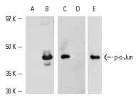
Western blot analysis of c-Jun activation in whole cell lysates from control (A) and anisomycin-treated (B) NIH/3T3 cells and phorbol ester-induced A-431 nuclear extracts (C,D,E). Antibodies tested include p-c-Jun (KM-1): sc-822 (A,B,C) and p-c-Jun (KM-1): sc-822 blocked with its cognate (D) or an unrelated peptide (E).
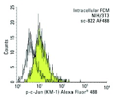
p-c-Jun (KM-1) Alexa Fluor 488: sc-822 AF488. Intracellular FCM analysis of fixed and permeabilized NIH/3T3 cells. Black line histogram represents the isotype control, normal mouse IgG
1: sc-3890.
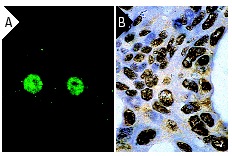
c-Jun (KM-1): sc-822. Immunofluorescence staining of methanol-fixed NIH/3T3 cells showing punctate nuclear staining of phosphorylated c-Jun (A). Immunoperoxidase staining of formalin-fixed, paraffin-embedded human colon carcinoma showing nuclear localization of activated c-Jun (B).
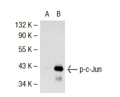
p-c-Jun (KM-1): sc-822. Western blot analysis of c-Jun phosphorylation in non-transfected: sc-117752 (A) and mouse c-Jun transfected: sc-125069 (B) 293T whole cell lysates.
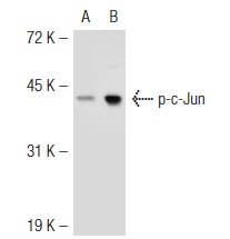
p-c-Jun (KM-1): sc-822. Western blot analysis of c-Jun phosphorylation in non-transfected: sc-117752 (A) and mouse c-Jun transfected: sc-125070 (B) 293T whole cell lysates.
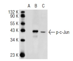
p-c-Jun (KM-1): sc-822. Western blot analysis of c-Jun phosphorylation in non-transfected CHO: sc-117750 (A), human c-Jun transfected CHO: sc-110019 (B) and NIH/3T3 (C) whole cell lysates.
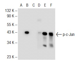
Western blot analysis of c-Jun phosphorylation in non-transfected: sc-117752 (A, D), untreated mouse c-Jun transfected: sc-125069 (B, E) and lambda protein phosphatase treated mouse c-Jun transfected: sc-125069 (C, F) 293T whole cell lysates. Antibodies tested include p-c-Jun (KM-1): sc-822 (A, B, C) and c-Jun (N): sc-45 (D, E, F).
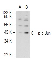
p-c-Jun (KM-1): sc-822. Western blot analysis of c-Jun phosphorylation in non-transfected: sc-117752 (A) and human c-Jun transfected: sc-159456 (B) 293T whole cell lysates.
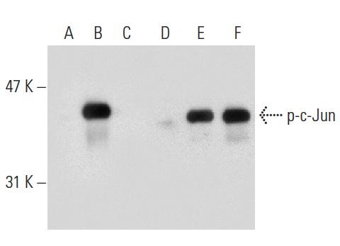
Western blot analysis of c-Jun phosphorylation in non-transfected: sc-117752 (A,D), untreated mouse c-Jun transfected: sc-125069 (B,E) and lambda protein phosphatase (sc-200312A) treated human c-Jun transfected: sc-125069 (C,F) 293T whole cell lysates. Antibodies tested include p-c-Jun (KM-1): sc-822 (A,B,C) and c-Jun (H-79): sc-1694 (D,E,F).
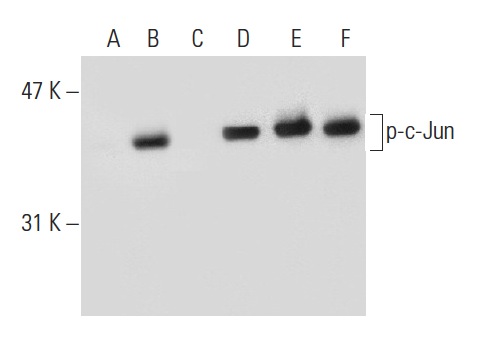
Western blot analysis of c-Jun phosphorylation in untreated (A,D), UV irradiated (B,E) and UV irradiated and lambda protein phosphatase (sc-200312A) treated (C,F) NIH/3T3 whole cell lysates. Antibodies tested include p-c-Jun (KM-1): sc-822 (A,B,C) and c-Jun (H-79): sc-1694 (D,E,F).
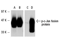
Western blot analysis of human recombinant c-Jun (A,C) and human recombinant c-Jun phosphorylated by human recombinant JNK1 (B,D) fusion proteins. Antibodies tested include c-Jun (N): sc-45 (A,B) and p-c-Jun (KM-1): sc-822 (C,D).
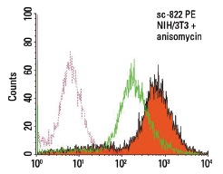
Anisomycin: sc-3524. Intracellular FCM analysis of Anisomycin (sc-3524) induced c-Jun serine-63 phosphorylation: Pink dotted lines represent mouse IgG1 PE stained Anisomycin (sc-3524) treated NIH/3T3 cells. Green line histograms represent untreated NIH/3T3 cells. Solid orange histograms represent anisomycin treated NIH/3T3 cells. p-c-Jun (KM-1) PE: sc-822 PE.


