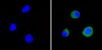
Immunofluorescent analysis of LFA3 (green) showing staining in the cytoplasm of Raji cells (right) compared to a negative control without primary antibody (left). Formalin-fixed cells were permeabilized with 0.1% Triton X-100 in TBS for 5-10 minutes and blocked with 3% BSA-PBS for 30 minutes at room temperature. Cells were probed with a LFA3 monoclonal antibody (ab171087) in 3% BSA-PBS at a dilution of 1:20 and incubated overnight at 4ºC in a humidified chamber. Cells were washed with PBST and incubated with a DyLight-conjugated secondary antibody in PBS at room temperature in the dark. F-actin (red) was stained with a fluorescent red phalloidin and nuclei (blue) were stained with Hoechst or DAPI. Images were taken at a magnification of 60x.
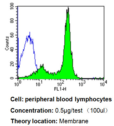
Flow cytometry analysis of LFA3 showing positive staining in the membrane of PBMC cells compared to an isotype control (blue). Human blood was collected, combined with a hydrophilic polysaccharide, centrifuged, transferred to a conical tube and washed with PBS. 50 ul of cell solution was added to each tube at a dilution of 2x10^7 cells/ml, followed by the addition of 50 ul of isotype control and ab171087 (0.5 ug/test). Cells were incubated for 30 min at 4ºC and washed with a cell buffer, followed by incubation with a DyLight 488-conjugated goat anti-mouse IgG (H+L) secondary for 30 min at 4ºC in the dark. FACS analysis was performed using 400 ul of cell buffer.
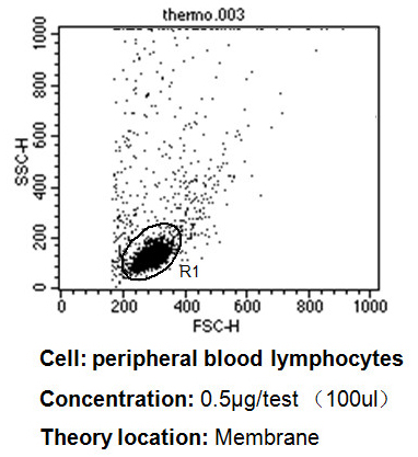
Flow cytometry analysis of LFA3 showing positive staining in the membrane of PBMC cells. Human blood was collected, combined with a hydrophilic polysaccharide, centrifuged, transferred to a conical tube and washed with PBS. 50 ul of cell solution was added to each tube at a dilution of 2x10^7 cells/ml, followed by the addition of 50 ul of isotype control and ab171087 (0.5 ug/test). Cells were incubated for 30 min at 4ºC and washed with a cell buffer, followed by incubation with a DyLight 488-conjugated goat anti-mouse IgG (H+L) secondary for 30 min at 4ºC in the dark. FACS analysis was performed using 400 ul of cell buffer.
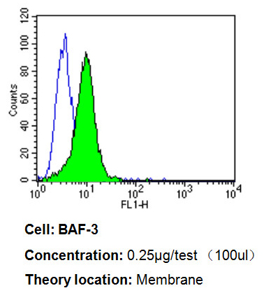
Flow cytometry analysis of LFA3 showing weakly positive staining in the membrane of BAF-3 cells compared to an isotype control (blue). Cells were harvested, adjusted to a concentration of 1-5x10^6 cells/ml, fixed with 2% paraformaldehyde, washed with PBS, and incubated with ab171087 (0.25 ug/test) for 60 min at room temperature. Cells were then blocked in a solution of 2% BSA-PBS for 30 min at room temperature, incubated for 40 min at room temperature in the dark using a Dylight 488-conjugated goat anti-mouse IgG (H+L) secondary antibody, and re-suspended in PBS for FACS analysis.
![All lanes : Anti-LFA3 antibody [TS2/9] (ab171087) at 1/100 dilutionLane 1 : Raji cell lysateLane 2 : Jurkat cell lysateLane 3 : BAF-3 cell lysateLysates/proteins at 25 µg per lane.](http://www.bioprodhub.com/system/product_images/ab_products/2/sub_3/15834_ab171087-201286-ab171087wb.jpg)
All lanes : Anti-LFA3 antibody [TS2/9] (ab171087) at 1/100 dilutionLane 1 : Raji cell lysateLane 2 : Jurkat cell lysateLane 3 : BAF-3 cell lysateLysates/proteins at 25 µg per lane.