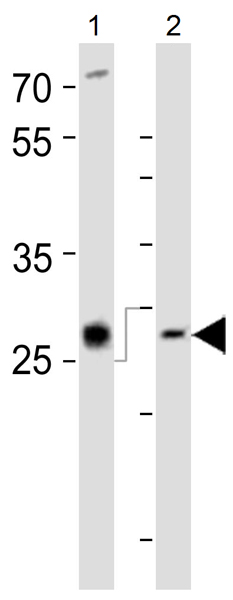
All lanes : Anti-LIF antibody (ab135629) at 1/1000 dilutionLane 1 : PC-12 cell lysateLane 2 : Mouse spleen tissue lysateLysates/proteins at 20 µg per lane.SecondaryHRP-conjugated goat anti-rabbit IgG (H+L) at 1/10000 dilution

Anti-LIF antibody (ab135629) at 1/1000 dilution + CEM cells lysate at 35 µgSecondaryHRP goat anti-rabbit (H+L) at 1/5000 dilution

All lanes : Anti-LIF antibody (ab135629) at 1/100 dilutionLane 1 : Nontransfected 293 cell lysates Lane 2 : lysate of 293 cells transiently transfected with the LIF gene Lysates/proteins at 2 µg per lane.

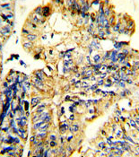
Immunohistochemical staining of LIF in Human colon carcinoma tissue sections (IHC-P - paraformaldehyde-fixed, paraffin-embedded sections) with ab135629 at a dilution of 1/25. Tissue was fixed with formaldehyde and blocked with 3% BSA for 0.5 hours at 38°C. Antigen retrieval was heat mediation with a citrate buffer (pH6). Samples were incubated with primary antibody (1/25) for 1 hour at 37°C. A HRP-conjugated goat anti-rabbit polyclonal (ready to use) was used as the secondary antibody.

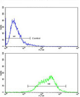
Flow cytometric analysis of CEM cells using ab135629 at 1/10 dilution staining LIF (bottom histogram) compared to a negative control cell (top histogram). FITC-conjugated goat-anti-rabbit secondary antibodies were used for the analysis.


