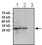
All lanes : Anti-Lin28 antibody (ab175352) at 1/1000 dilutionLane 1 : NCCIT whole cell lysateLane 2 : NTERA-2 whole cell lysateLane 3 : HeLa whole cell lysateLysates/proteins at 75 µg per lane.Secondarygoat anti-rabbit-HRP secondary antibody at 1/20000 dilution

Immunofluorescent analysis of formalin-fixed and permeabilized HeLa cells labeling Lin28 with ab175352 at 1/100 and incubated with a DyLight-conjugated secondary antibody. F-actin (red) was stained with a fluorescent phalloidin and nuclei (blue) were stained with DAPI.

Immunofluorescent analysis of fH9 embryonic stem cells labeling Lin28 with ab175352 at 1/200 and incubated with a fluorescently-conjugated secondary antibody. Nuclei (blue) were stained with DAPI.

Immunofluorescent analysis of HEL 11.4 induced IPS cells labeling Lin28 with ab175352 at 1/200 and incubated with a fluorescently-conjugated secondary antibody. Nuclei (blue) were stained with DAPI.
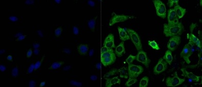
Immunofluorescent analysis of formalin-fixed and permeabilized HeLa cells (left image) and Human embryonal carcinoma NTERA-2 cells (right image) labeling Lin28 with ab175352 at 1/200 and and incubated with DyLight 488 goat-anti-rabbit IgG secondary antibody. Nuclei (blue) were stained with Hoechst 33342 dye.
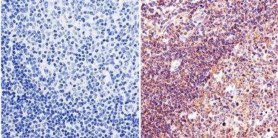
Immunohistochemical analysis of paraffin embedded Human tonsil tissue labeling Lin28 with ab175352 at 1/50.
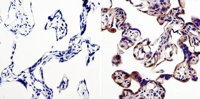
Immunohistochemical analysis of paraffin embedded Human placenta tissue labeling Lin28 with ab175352 at 1/50.
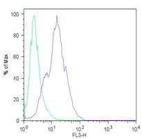
Flow Cytometrical analysis of H9 embryonic stem cells labeling Lin28 with ab175352 at 1/100 (right histogram) compared to rabbit IgGl (left histogram) and incubated with a fluorescently-conjugated secondary antibody.
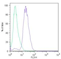
Flow Cytometrical analysis of HEL 11.4 induced IPS cells labeling Lin28 with ab175352 at 1/100 (right histogram) compared to rabbit IgGl (left histogram) and incubated with a fluorescently-conjugated secondary antibody.

Lane 2: Immunoprecipitation. ab175352 at 1/1000 labeling Lin28 immunoprecipitated from 750 μg of NCCIT whole cell lysate using ab175352 at 3μg.


