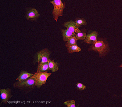
ICC/IF image of ab102959 stained DU145 cells. The cells were 4% formaldehyde fixed (10 min) and then incubated in 1%BSA / 10% normal goat serum / 0.3M glycine in 0.1% PBS-Tween for 1h to permeabilise the cells and block non-specific protein-protein interactions. The cells were then incubated with the antibody ab102959 at 5µg/ml overnight at +4°C. The secondary antibody (green) was DyLight® 488 goat anti- rabbit (ab96899) IgG (H+L) used at a 1/250 dilution for 1h. Alexa Fluor® 594 WGA was used to label plasma membranes (red) at a 1/200 dilution for 1h. DAPI was used to stain the cell nuclei (blue) at a concentration of 1.43µM.

Anti-Mitoferrin1 antibody (ab102959) at 1/100 dilution + MDA-MB231 cell line lysate at 35 µg

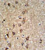
IHC analysis in formalin fixed and paraffin embedded Human brain tissue followed by peroxidase conjugation of the secondary antibody and DAB staining, using ab102959 at a dilution of 1/50.


