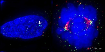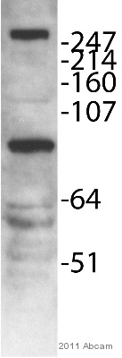
ab75339 staining MLL5 in HeLa cells by Immunocytochemistry/ Immunoflourescence. Cells were fixed in Methanol and permeabilized in PHEM/0.5% Triton X-100 prior to blocking in 2.5% BSA for 1 hour at 21°C. The primary antibody was diluted 1/100 and incubated with the sample for 1 hour at 21°C. The secondary antibody was FITC-conjugated Donkey anti-Rabbit polyclonal, diluted 1/150. DNA (DAPI) stain in blue, anti-Aurora A antibody in red, anti-MLL5 in green. Left cell shows centrosomal staining in interphase (arrow head). Right cell shows centrosomal staining in mitotic cell. Centrosomal localisation of MLL5 was shown in Liu et al (2010) PMID:20439461See Abreview

