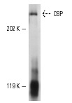
CBP (A-22): sc-369. Western blot analysis of CBP expression in KNRK nuclear extract.

ChIP analysis of transcription factor binding to the Interferon β promoter before (A) and six hours after (B) Sendai virus infection of HeLa cells. Antibodies tested included NFκB p65 (A): sc-109, ATF-2 (N-96): sc-6233, GCN5 (N-18): sc-6303, Pol II (H-224): sc-9001, TFIID (TBP)(SI-1): sc-273 and CBP (A-22): sc-369. Data kindly provided by G. Mosialos.
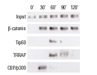
ChIP analysis of coactivator recruitment on Cyclin D2 promoter in C2C12 cells treated with LiCl and serum. Antibodies tested include β-catenin (H-102): sc-7199, β-catenin (C-18): sc-1496, β-catenin (E-5): sc-7963, Tip60 (N-17): sc-5725, TRRAP (T-17): sc-5405, TRRAP (Y-18): sc-12375, TRRAP (F-20): sc-12376, TRRAP (H-300): sc-11411, CBP (A-22): sc-369, CBP (C-20): sc-583, CBP (451): sc-1211, CPB (C-1): sc-7300, p300 (H-272): sc-8981, p300 (N-15): sc-584 and p300 (C-20): sc-585. Data kindly provided by M.G. Rosenfeld and reproduced with permission from Kioussi et al., Cell 2002, 111: 673-685.
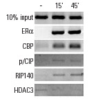
ChIP analysis of p52 promoter occupancy in murine p19 cells in response to activation by estradiol (E2). Antibodies tested include ERα (MC-20): sc-542, ERα (H-184): sc-7207, CBP (A-22): sc-369, CBP (C-1): sc-7300, p/CIP (F2): sc-5305, p/CIP (M-397): sc-9119, RIP140 (H-300): sc-8997 and HDAC3 (N-19): sc-11417. Data kindly provided by M.G. Rosenfeld and reproduced with permission from Perissi et al., Cell 2004, 116: 511-526.
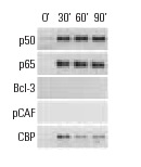
ChIP analysis of cofactor occupancy dynamics on the ICAM1 promoter in 293 cells in response to IL-1β treatment. Antibodies tested include NFκB p50 (C-19): sc-1190, NFκB p50 (E-10): sc-8414, NFκB p50 (H-119): sc-7178, NFκB p65 (C-20): sc-372, NFκB p65 (A): sc-109, NFκB p65 (H-286): sc-7151, Bcl-3 (C-14): sc-185, Bcl-3 (H-146): sc-13038, PCAF (C-16): sc-6300, PCAF (H-369): sc-8999, CBP (A-22): sc-369, CBP (C-1): sc-7300, CBP (C-20): sc-583, CBP (451): sc-1211. Data kindly provided by M.G. Rosenfeld and reproduced with permission from Baek et al., Cell 2002, 110: 55-67.
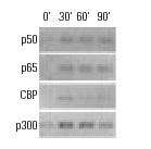
ChIP analysis of cofactor occupancy dynamics on the IL-8 promoter in 293 cells in response to IL-1β treatment. Antibodies tested include NFκB p50 (C-19): sc-1190, NFκB p50 (E-10): sc-8414, NFκB p50 (H-119): sc-7178, NFκB p65 (C-20): sc-372, NFκB p65 (A): sc-109, NFκB p65 (H-286): sc-7151, CBP (A-22): sc-369, CBP (C-1): sc-7300, CBP (C-20): sc-583, CBP (451): sc-1211, p300 (C-20): sc-sc-585, p300 (N-15): sc-584, p300 (H-272): sc-8981. Data kindly provided by M.G. Rosenfeld and reproduced with permission from Baek et al., Cell 2002, 110: 55-67.
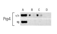
ChIP analysis of in vivo binding of RORα and its recruitment of coactivators to RORα-responsive promoters in freshly dissected cerebella derived from wild type (+/+) and staggerer (Sg) mice. Control Input (A). Antibodies used included RORα (C-16): sc-6062 (B), TIP60 (N-17): sc-5725 (C), CBP (A-22): sc-369 and CBP (C-20): sc-583 (D). Data kindly provided by M.G. Rosenfeld and reproduced wtih permission from Gold et al., Neuron 2003, 40: 1119-1131.

ChIP analysis of in vivo binding of RORα and its recruitment of coactivators to RORα-responsive promoters in freshly dissected cerebella derived from wild type (+/+) and staggerer (Sg) mice. Control Input (A). Antibodies used included RORα (C-16): sc-6062 (B), TIP60 (N-17): sc-5725 (C), CBP (A-22): sc-369 and CBP (C-20): sc-583 (D). Data kindly provided by M.G. Rosenfeld and reproduced wtih permission from Gold et al., Neuron 2003, 40: 1119-1131.
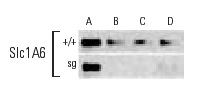
ChIP analysis of in vivo binding of RORα and its recruitment of coactivators to RORα-responsive promoters in freshly dissected cerebella derived from wild type (+/+) and staggerer (Sg) mice. Control Input (A). Antibodies used included RORα (C-16): sc-6062 (B), TIP60 (N-17): sc-5725 (C), CBP (A-22): sc-369 and CBP (C-20): sc-583 (D). Data kindly provided by M.G. Rosenfeld and reproduced wtih permission from Gold et al., Neuron 2003, 40: 1119-1131.

ChIP analysis of in vivo binding of RORα and its recruitment of coactivators to RORα-responsive promoters in freshly dissected cerebella derived from wild type (+/+) and staggerer (Sg) mice. Control Input (A). Antibodies used included RORα (C-16): sc-6062 (B), TIP60 (N-17): sc-5725 (C), CBP (A-22): sc-369 and CBP (C-20): sc-583 (D). Data kindly provided by M.G. Rosenfeld and reproduced wtih permission from Gold et al., Neuron 2003, 40: 1119-1131.
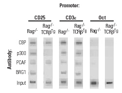
ChIP analysis of recruitment of CBP, p300, PCA and BRG1 to the IL-2α (CD25) promoter in vivo. Antibodies tested include CBP (A-22): sc-369, p300 (N-15): sc-584, PCAF (H-369): sc-8999, and Brg-1 (H-88): sc-10768. CD3ε and Oct-2 promoter regions were employed as positive and negative controls, respectively. DNA was isolated from Rag
-/- thymocytes or Rag
-/- thymocytes expressing TCRβ. Data kindly provided by J. Imbert and reproduced from Yeh, J-H., et al., Nucleic Acids Research, 2002, 30: 1944-1951, with permission from Oxford University Press.

ChIP analysis of
in vivo binding of RORα and its recruitment of coactivators to RORα-responsive promoters in freshly dissected cerebella derived from wild type (+/+) and staggerer (Sg) mice. Control Input (A). Antibodies used included RORa (C-16): sc-6062 (B), TIP60 (N-17): sc-5725 (C), CBP (A-22): sc-369 and CBP (C-20): sc-583 (D). Data kindly provided by M.G. Rosenfeld and reproduced wtih permission from Gold
et al., Neuron 2003, 40: 1119-1131.
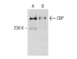
CBP (A-22): sc-369. Western blot analysis of CBP expression in KNRK (A) and Jurkat (B) nuclear extracts.


