Anti-NADPH oxidase 4 antibody [UOTR1B492]
| Name | Anti-NADPH oxidase 4 antibody [UOTR1B492] |
|---|---|
| Supplier | Abcam |
| Catalog | ab109225 |
| Host | Rabbit |
| Clonality | Monoclonal |
| Isotype | IgG |
| Clone | UOTR1B492 |
| Applications | WB IP IHC-P ICC/IF |
| Species Reactivities | Mouse, Rat, Dog, Human |
| Antigen | Synthetic peptide (the amino acid sequence is considered to be commercially sensitive) corresponding to Human NADPH oxidase 4 aa 500-600 |
| Blocking Peptide | NADPH oxidase 4 peptide |
| Description | Rabbit Monoclonal |
| Gene | NOX4 |
| Conjugate | Unconjugated |
| Supplier Page | Shop |
Product images
![Anti-NADPH oxidase 4 antibody [UOTR1B492] (ab109225) at 1/10000 dilution (purified) + JAR cell lysate at 10 µgSecondaryHRP goat anti-rabbit (H+L) at 1/1000 dilution](http://www.bioprodhub.com/system/product_images/ab_products/2/sub_3/27310_ab109225-237251-109225-WB-2.jpg) Anti-NADPH oxidase 4 antibody [UOTR1B492] (ab109225) at 1/10000 dilution (purified) + JAR cell lysate at 10 µgSecondaryHRP goat anti-rabbit (H+L) at 1/1000 dilution
Anti-NADPH oxidase 4 antibody [UOTR1B492] (ab109225) at 1/10000 dilution (purified) + JAR cell lysate at 10 µgSecondaryHRP goat anti-rabbit (H+L) at 1/1000 dilution
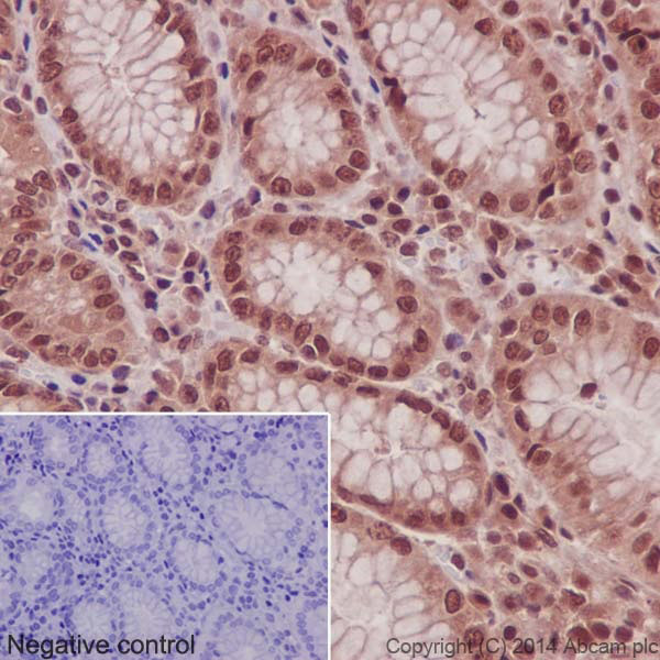 Immunohistochemical staining of paraffin embedded human stomach with purified ab109225 at a dilution of 1/500. A HRP goat anti-rabbit (ab97051) was used as the secondary antibody at a dilution of 1/500 and the sample was counter-stained with hematoxylin. Antigen retrieval was perfomed using Tris-EDTA buffer, pH 9.0. PBS was used instead of the primary antibody as the negative control, and is shown in the inset.
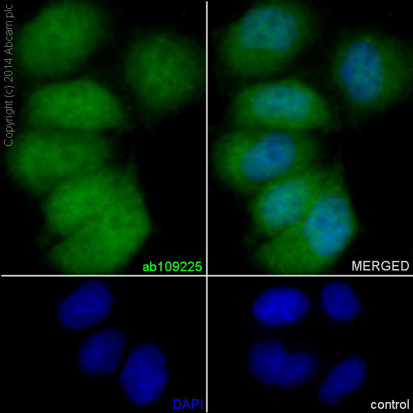 Immunofluorescent staining of HeLa cells (fixed in 4% PFA, permeabilized with 0.1% Triton X 100) using purified ab109225 at a dilution of 1/200. An Alexa Fluor® 488 goat anti-rabbit antibody was used as the secondary at a dilution of 1/500 and the cells were counter stained with DAPI. The negative control is shown in the bottom right hand panel - for the negative control, Alex Fluor® 594 goat anti-mouse was used at a dilution of 1/500.
![Anti-NADPH oxidase 4 antibody [UOTR1B492] (ab109225) at 1/2000 dilution (purified) + Human fetal kidney at 10 µgSecondaryHRP anti-rabbit, specific to the non reducded form of IgG at 1/1000 dilution](http://www.bioprodhub.com/system/product_images/ab_products/2/sub_3/27314_ab109225-237246-109225-WB-1.jpg) Anti-NADPH oxidase 4 antibody [UOTR1B492] (ab109225) at 1/2000 dilution (purified) + Human fetal kidney at 10 µgSecondaryHRP anti-rabbit, specific to the non reducded form of IgG at 1/1000 dilution
Anti-NADPH oxidase 4 antibody [UOTR1B492] (ab109225) at 1/2000 dilution (purified) + Human fetal kidney at 10 µgSecondaryHRP anti-rabbit, specific to the non reducded form of IgG at 1/1000 dilution
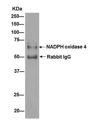 ab109225 (purified) at 1/30 immunoprecipitating NADPH oxidase 4 in HEK293. For western blotting, a HRP-conjugated anti-rabbit antibody was used as the secondary antibody (1/1000).Blocking buffer and concentration: 5% NFDM/TBST.Diluting buffer and concentration: 5% NFDM /TBST.
![All lanes : Anti-NADPH oxidase 4 antibody [UOTR1B492] (ab109225) at 1/1000 dilution (unpurified)Lane 1 : Fetal kidney lysateLane 2 : U87-MG lysateLane 3 : 293T lysateLane 4 : JAR lysates Lysates/proteins at 10 µg per lane.SecondaryStandard HRP labelled goat anti-rabbit at 1/2000 dilution](http://www.bioprodhub.com/system/product_images/ab_products/2/sub_3/27316_NOX4-Primary-antibodies-ab109225-1.jpg) All lanes : Anti-NADPH oxidase 4 antibody [UOTR1B492] (ab109225) at 1/1000 dilution (unpurified)Lane 1 : Fetal kidney lysateLane 2 : U87-MG lysateLane 3 : 293T lysateLane 4 : JAR lysates Lysates/proteins at 10 µg per lane.SecondaryStandard HRP labelled goat anti-rabbit at 1/2000 dilution
All lanes : Anti-NADPH oxidase 4 antibody [UOTR1B492] (ab109225) at 1/1000 dilution (unpurified)Lane 1 : Fetal kidney lysateLane 2 : U87-MG lysateLane 3 : 293T lysateLane 4 : JAR lysates Lysates/proteins at 10 µg per lane.SecondaryStandard HRP labelled goat anti-rabbit at 1/2000 dilution
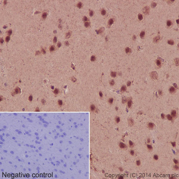
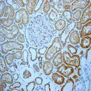 Immunohistochemical analysis of paraffin-embedded Human kidney tissue using unpurified ab109225.
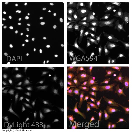 ICC/IF image of unpurified ab109255 stained HeLa cells. The cells were 4% formaldehyde fixed (10 min) and then incubated in 1%BSA / 10% normal goat serum / 0.3M glycine in 0.1% PBS-Tween for 1h to permeabilise the cells and block non-specific protein-protein interactions. The cells were then incubated with the antibody (ab109225, 5µg/ml) overnight at +4°C. The secondary antibody (green) was ab96899, DyLight® 488 goat anti-rabbit IgG (H+L) used at a 1/250 dilution for 1h. Alexa Fluor® 594 WGA was used to label plasma membranes (red) at a 1/200 dilution for 1h. DAPI was used to stain the cell nuclei (blue) at a concentration of 1.43µM.
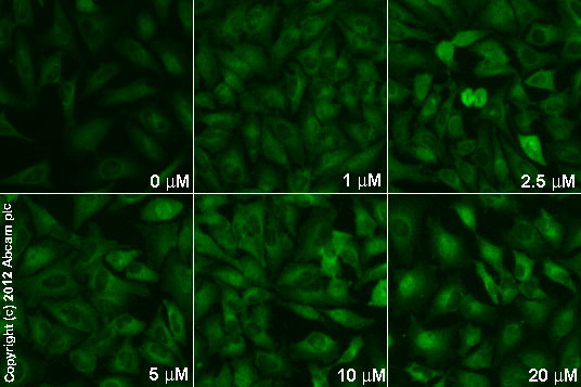 Unpurified ab109225 staining Nox4 in HeLa cells treated with (-)-cannabidiol (ab120448), by ICC/IF. Increase in Nox4 expression correlates with increased concentration of (-)-cannabidiol, as described in literature.The cells were incubated at 37°C for 6h in media containing different concentrations of ab120448 ((-)-cannabidio) in DMSO, fixed with 4% formaldehyde for 10 minutes at room temperature and blocked with PBS containing 10% goat serum, 0.3 M glycine, 1% BSA and 0.1% tween for 2h at room temperature. Staining of the treated cells with ab109225 (5 µg/ml) was performed overnight at 4°C in PBS containing 1% BSA and 0.1% tween. A DyLight 488 goat anti-rabbit polyclonal antibody (ab96899) at 1/250 dilution was used as the secondary antibody.
Product References
Iron chelation by deferoxamine prevents renal interstitial fibrosis in mice with - Iron chelation by deferoxamine prevents renal interstitial fibrosis in mice with
Ikeda Y, Ozono I, Tajima S, Imao M, Horinouchi Y, Izawa-Ishizawa Y, Kihira Y, Miyamoto L, Ishizawa K, Tsuchiya K, Tamaki T. PLoS One. 2014 Feb 19;9(2):e89355.
Endothelial dysfunction in adipose triglyceride lipase deficiency. - Endothelial dysfunction in adipose triglyceride lipase deficiency.
Schrammel A, Mussbacher M, Wolkart G, Stessel H, Pail K, Winkler S, Schweiger M, Haemmerle G, Al Zoughbi W, Hofler G, Lametschwandtner A, Zechner R, Mayer B. Biochim Biophys Acta. 2014 Jun;1841(6):906-17.
Macrophage migration inhibitory factor antagonizes pressure overload-induced - Macrophage migration inhibitory factor antagonizes pressure overload-induced
Koga K, Kenessey A, Ojamaa K. Am J Physiol Heart Circ Physiol. 2013 Jan 15;304(2):H282-93. doi:
Angiotensin-converting enzyme 2 priming enhances the function of endothelial - Angiotensin-converting enzyme 2 priming enhances the function of endothelial
Chen J, Xiao X, Chen S, Zhang C, Chen J, Yi D, Shenoy V, Raizada MK, Zhao B, Chen Y. Hypertension. 2013 Mar;61(3):681-9.
TRAF3IP2 mediates interleukin-18-induced cardiac fibroblast migration and - TRAF3IP2 mediates interleukin-18-induced cardiac fibroblast migration and
Valente AJ, Sakamuri SS, Siddesha JM, Yoshida T, Gardner JD, Prabhu R, Siebenlist U, Chandrasekar B. Cell Signal. 2013 Nov;25(11):2176-84.
Role of renal DJ-1 in the pathogenesis of hypertension associated with increased - Role of renal DJ-1 in the pathogenesis of hypertension associated with increased
Cuevas S, Zhang Y, Yang Y, Escano C, Asico L, Jones JE, Armando I, Jose PA. Hypertension. 2012 Feb;59(2):446-52.
Nox4 is a novel inducible source of reactive oxygen species in monocytes and - Nox4 is a novel inducible source of reactive oxygen species in monocytes and
Lee CF, Qiao M, Schroder K, Zhao Q, Asmis R. Circ Res. 2010 May 14;106(9):1489-97.