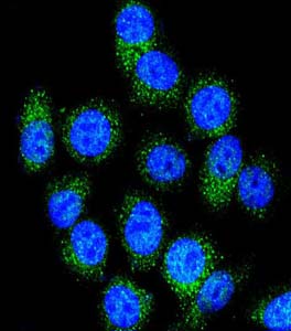
Confocal immunofluorescence analysis of Hela cells labeling NAT1 with ab174969 at 1/10 dilution, followed by Alexa Fluor® 488-conjugated goat anti-rabbit lgG (green). DAPI was used to stain the cell nuclear (blue).
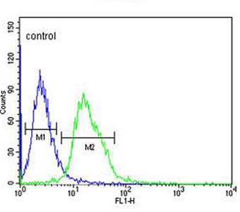
Flow cytometric analysis of CEM cells (bottom histogram) labeling NAT1 with ab174969 at 1/10 dilution compared to a negative control cell (top histogram). FITC-conjugated goat-anti-rabbit secondary antibodies were used for the analysis
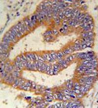
Immunohistochemical analysis of formalin fixed and paraffin embedded Human colon carcinoma labeling NAT1 with ab174969 at 1/50 dilution, followed by peroxidase conjugation of the secondary antibody and DAB staining.
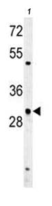
Anti-NAT1 antibody - C-terminal (ab174969) at 1/100 dilution + CEM cell line lysate at 35 µgdeveloped using the ECL technique