![Anti-NDUFS6 antibody [EPR15957] (ab195807) at 1/10000 dilution + Human fetal kidney lysate at 20 µgSecondaryAnti-Rabbit IgG (HRP), specific to the non-reduced form of IgG at 1/1000 dilution](http://www.bioprodhub.com/system/product_images/ab_products/2/sub_3/28664_ab195807-239871-ab195807.jpg)
Anti-NDUFS6 antibody [EPR15957] (ab195807) at 1/10000 dilution + Human fetal kidney lysate at 20 µgSecondaryAnti-Rabbit IgG (HRP), specific to the non-reduced form of IgG at 1/1000 dilution
![Anti-NDUFS6 antibody [EPR15957] (ab195807) at 1/10000 dilution + Human fetal heart at 20 µgSecondaryAnti-Rabbit IgG (HRP), specific to the non-reduced form of IgG at 1/1000 dilution](http://www.bioprodhub.com/system/product_images/ab_products/2/sub_3/28665_ab195807-239872-ab195807b.jpg)
Anti-NDUFS6 antibody [EPR15957] (ab195807) at 1/10000 dilution + Human fetal heart at 20 µgSecondaryAnti-Rabbit IgG (HRP), specific to the non-reduced form of IgG at 1/1000 dilution
![Anti-NDUFS6 antibody [EPR15957] (ab195807) at 1/10000 dilution + Human skeletal muscle lysate at 20 µgSecondaryAnti-Rabbit IgG (HRP), specific to the non-reduced form of IgG at 1/1000 dilution](http://www.bioprodhub.com/system/product_images/ab_products/2/sub_3/28666_ab195807-239873-ab195807c.jpg)
Anti-NDUFS6 antibody [EPR15957] (ab195807) at 1/10000 dilution + Human skeletal muscle lysate at 20 µgSecondaryAnti-Rabbit IgG (HRP), specific to the non-reduced form of IgG at 1/1000 dilution
![All lanes : Anti-NDUFS6 antibody [EPR15957] (ab195807) at 1/1000 dilutionLane 1 : Mouse brain lysateLane 2 : Mouse heart lysateLane 3 : Rat brain lysateLane 4 : Rat heart lysateLane 5 : Rat kidney lysateLysates/proteins at 10 µg per lane.SecondaryAnti-Rabbit IgG (HRP), specific to the non-reduced form of IgG at 1/1000 dilution](http://www.bioprodhub.com/system/product_images/ab_products/2/sub_3/28667_ab195807-239870-ab195807d.jpg)
All lanes : Anti-NDUFS6 antibody [EPR15957] (ab195807) at 1/1000 dilutionLane 1 : Mouse brain lysateLane 2 : Mouse heart lysateLane 3 : Rat brain lysateLane 4 : Rat heart lysateLane 5 : Rat kidney lysateLysates/proteins at 10 µg per lane.SecondaryAnti-Rabbit IgG (HRP), specific to the non-reduced form of IgG at 1/1000 dilution

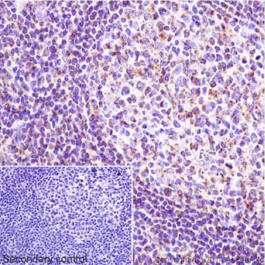
Immunohistochemical analysis of paraffin-embedded Human tonsil tissue labeling NDUFS6 with ab195807 at 1/500. Secondary antibody: Goat Anti-Rabbit IgG H&L (HRP) (ab97051) at 1/500. Inset image: negative control obtained using PBS instead of ab195807 and secondary antibody only.Note: Cytoplasm staining on human tonsil tissue was observed.

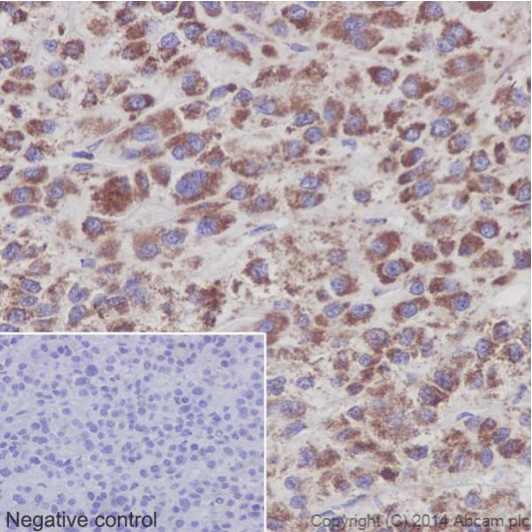
Immunohistochemical analysis of paraffin-embedded Human hepatocellular carcinoma tissue labeling NDUFS6 with ab195807 at 1/500. Secondary antibody: Goat Anti-Rabbit IgG H&L (HRP) (ab97051) at 1/500.Inset image: negative control obtained using PBS instead of ab195807 and secondary antibody only.Note: Cytoplasm staining on human hepatocellular carcinoma tissue was observed.

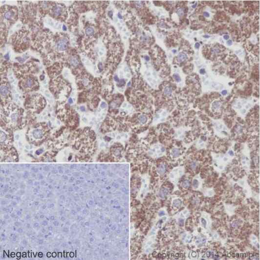
Immunohistochemical analysis of paraffin-embedded Rat liver tissue labeling NDUFS6 with ab195807 at 1/500. Secondary antibody: Goat Anti-Rabbit IgG H&L (HRP) (ab97051) at 1/500.Inset image: negative control obtained using PBS instead of ab195807 and secondary antibody only.Note: Cytoplasm staining on rat liver tissue was observed.

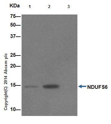
Immunoprecipitation analysis of Human fetal kidney lysate labeling NDUFS6 using ab195807 at 1/30 dilution (Lane 2).Lane 3: IP using Rabbit monoclonal IgG (ab172730) ) instead of ab195807 in Human fetal kidney lysates.Lane 1: Input: 10 μg Human fetal kidney lysates.Subsequent WB detection was performed using ab195807 at 1/1000 dilution.An Anti-Rabbit IgG (HRP), specific to the non-reduced form of IgG at 1/1500 was used as secondary antibody.
![Anti-NDUFS6 antibody [EPR15957] (ab195807) at 1/10000 dilution + Human fetal kidney lysate at 20 µgSecondaryAnti-Rabbit IgG (HRP), specific to the non-reduced form of IgG at 1/1000 dilution](http://www.bioprodhub.com/system/product_images/ab_products/2/sub_3/28664_ab195807-239871-ab195807.jpg)
![Anti-NDUFS6 antibody [EPR15957] (ab195807) at 1/10000 dilution + Human fetal heart at 20 µgSecondaryAnti-Rabbit IgG (HRP), specific to the non-reduced form of IgG at 1/1000 dilution](http://www.bioprodhub.com/system/product_images/ab_products/2/sub_3/28665_ab195807-239872-ab195807b.jpg)
![Anti-NDUFS6 antibody [EPR15957] (ab195807) at 1/10000 dilution + Human skeletal muscle lysate at 20 µgSecondaryAnti-Rabbit IgG (HRP), specific to the non-reduced form of IgG at 1/1000 dilution](http://www.bioprodhub.com/system/product_images/ab_products/2/sub_3/28666_ab195807-239873-ab195807c.jpg)
![All lanes : Anti-NDUFS6 antibody [EPR15957] (ab195807) at 1/1000 dilutionLane 1 : Mouse brain lysateLane 2 : Mouse heart lysateLane 3 : Rat brain lysateLane 4 : Rat heart lysateLane 5 : Rat kidney lysateLysates/proteins at 10 µg per lane.SecondaryAnti-Rabbit IgG (HRP), specific to the non-reduced form of IgG at 1/1000 dilution](http://www.bioprodhub.com/system/product_images/ab_products/2/sub_3/28667_ab195807-239870-ab195807d.jpg)


