Anti-Neurogenin3 antibody
| Name | Anti-Neurogenin3 antibody |
|---|---|
| Supplier | Abcam |
| Catalog | ab38548 |
| Prices | $385.00 |
| Sizes | 400 µl |
| Host | Rabbit |
| Clonality | Polyclonal |
| Isotype | IgG |
| Applications | ICC/IF ICC/IF WB IHC-P |
| Species Reactivities | Mouse, Human |
| Antigen | Synthetic peptide corresponding to Human Neurogenin3 aa 40-69 (N terminal) conjugated to Keyhole Limpet Haemocyanin (KLH) |
| Description | Rabbit Polyclonal |
| Gene | NEUROG3 |
| Conjugate | Unconjugated |
| Supplier Page | Shop |
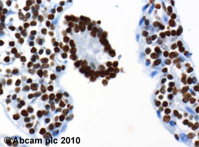
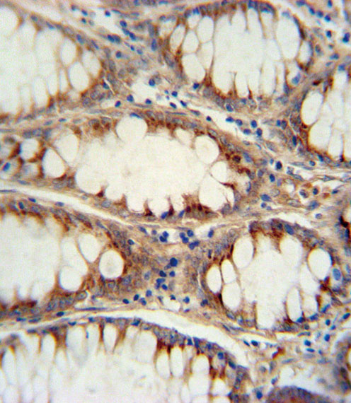
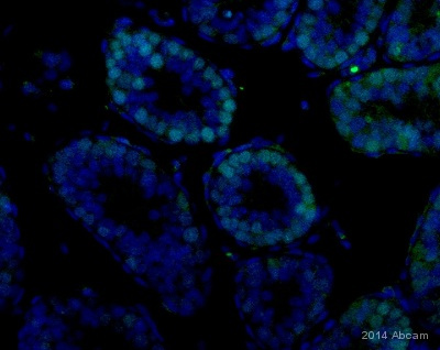
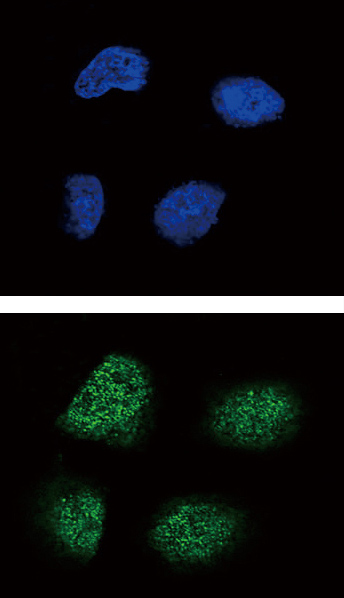
Product images
Product References
Neonatal diabetes and congenital malabsorptive diarrhea attributable to a novel - Neonatal diabetes and congenital malabsorptive diarrhea attributable to a novel
Pinney SE, Oliver-Krasinski J, Ernst L, Hughes N, Patel P, Stoffers DA, Russo P, De Leon DD. J Clin Endocrinol Metab. 2011 Jul;96(7):1960-5.
Gene expression profiling of a mouse model of pancreatic islet dysmorphogenesis. - Gene expression profiling of a mouse model of pancreatic islet dysmorphogenesis.
Wilding Crawford L, Tweedie Ables E, Oh YA, Boone B, Levy S, Gannon M. PLoS One. 2008 Feb 20;3(2):e1611.


