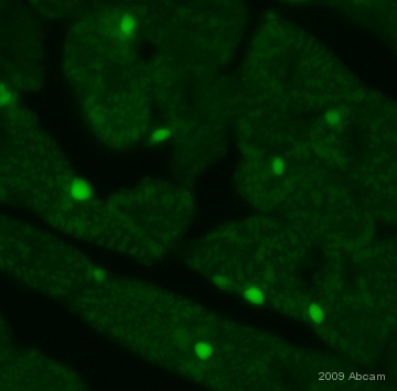
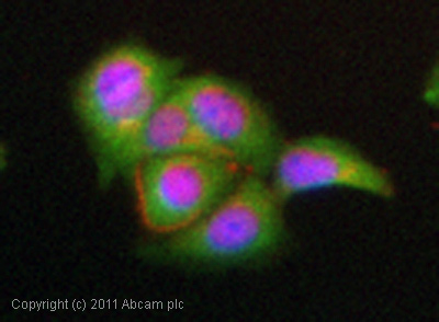
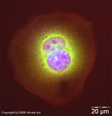
Anti-NF-kB p65 antibody
| Name | Anti-NF-kB p65 antibody |
|---|---|
| Supplier | Abcam |
| Catalog | ab16502 |
| Prices | $403.00 |
| Sizes | 100 µg |
| Host | Rabbit |
| Clonality | Polyclonal |
| Isotype | IgG |
| Applications | IHC-F ICC/IF ICC/IF IHC-P WB IP FC |
| Species Reactivities | Mouse, Rat, Chicken, Human, Deer |
| Antigen | Synthetic peptide corresponding to Human NF-kB p65 aa 500 to the C-terminus (C terminal) conjugated to Keyhole Limpet Haemocyanin (KLH) |
| Blocking Peptide | Human NF-kB p65 peptide |
| Description | Rabbit Polyclonal |
| Gene | RELA |
| Conjugate | Unconjugated |
| Supplier Page | Shop |
Product images
Product References
Clearance of senescent hepatocytes in a neoplastic-prone microenvironment delays - Clearance of senescent hepatocytes in a neoplastic-prone microenvironment delays
Marongiu F, Serra MP, Sini M, Angius F, Laconi E. Aging (Albany NY). 2014 Jan;6(1):26-34.
Overexpression of heat shock protein 72 attenuates NF-kappaB activation using a - Overexpression of heat shock protein 72 attenuates NF-kappaB activation using a
Sheppard PW, Sun X, Khammash M, Giffard RG. PLoS Comput Biol. 2014 Feb 6;10(2):e1003471.
CD31 is a key coinhibitory receptor in the development of immunogenic dendritic - CD31 is a key coinhibitory receptor in the development of immunogenic dendritic
Clement M, Fornasa G, Guedj K, Ben Mkaddem S, Gaston AT, Khallou-Laschet J, Morvan M, Nicoletti A, Caligiuri G. Proc Natl Acad Sci U S A. 2014 Mar 25;111(12):E1101-10. doi:
Nrf2 upregulates ATP binding cassette transporter expression and activity at the - Nrf2 upregulates ATP binding cassette transporter expression and activity at the
Wang X, Campos CR, Peart JC, Smith LK, Boni JL, Cannon RE, Miller DS. J Neurosci. 2014 Jun 18;34(25):8585-93.
A gain-of-function mouse model identifies PRMT6 as a NF-kappaB coactivator. - A gain-of-function mouse model identifies PRMT6 as a NF-kappaB coactivator.
Di Lorenzo A, Yang Y, Macaluso M, Bedford MT. Nucleic Acids Res. 2014 Jul;42(13):8297-309.
Comparative analysis of the cytotoxic effects of okadaic acid-group toxins on - Comparative analysis of the cytotoxic effects of okadaic acid-group toxins on
Ferron PJ, Hogeveen K, Fessard V, Le Hegarat L. Mar Drugs. 2014 Aug 21;12(8):4616-34.
MCPIP1 contributes to the toxicity of proteasome inhibitor MG-132 in HeLa cells - MCPIP1 contributes to the toxicity of proteasome inhibitor MG-132 in HeLa cells
Skalniak L, Dziendziel M, Jura J. Mol Cell Biochem. 2014 Oct;395(1-2):253-63.
Low-dose arsenic induces chemotherapy protection via p53/NF-kappaB-mediated - Low-dose arsenic induces chemotherapy protection via p53/NF-kappaB-mediated
Ganapathy S, Xiao S, Seo SJ, Lall R, Yang M, Xu T, Su H, Shadfan M, Ha CS, Yuan ZM. Oncogene. 2014 Mar 13;33(11):1359-66.
Induction of cytopathogenicity in human glioblastoma cells by chikungunya virus. - Induction of cytopathogenicity in human glioblastoma cells by chikungunya virus.
Abraham R, Mudaliar P, Padmanabhan A, Sreekumar E. PLoS One. 2013 Sep 25;8(9):e75854.
Ezrin-radixin-moesin-binding phosphoprotein 50 (EBP50) and nuclear factor-kappaB - Ezrin-radixin-moesin-binding phosphoprotein 50 (EBP50) and nuclear factor-kappaB
Leslie KL, Song GJ, Barrick S, Wehbi VL, Vilardaga JP, Bauer PM, Bisello A. J Biol Chem. 2013 Dec 20;288(51):36426-36.
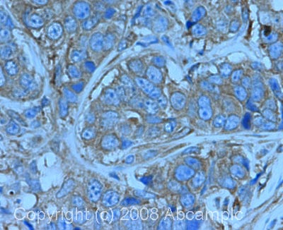

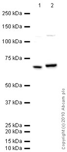
![NF-kB p65 was immunoprecipitated using 0.5mg Hela whole cell extract, 5µg of Rabbit polyclonal to NFkB p65 and 50µl of protein G magnetic beads (+). No antibody was added to the control (-). The antibody was incubated under agitation with Protein G beads for 10min, Hela whole cell extract lysate diluted in RIPA buffer was added to each sample and incubated for a further 10min under agitation.Proteins were eluted by addition of 40µl SDS loading buffer and incubated for 10min at 70oC; 10µl of each sample was separated on a SDS PAGE gel, transferred to a nitrocellulose membrane, blocked with 5% BSA and probed with ab16502.Secondary: Mouse monoclonal [SB62a] Secondary Antibody Anti-Rabbit HRP (IgG light chain) (ab99697).Band: 68kDa: NFkB p65](http://www.bioprodhub.com/system/product_images/ab_products/2/sub_3/29718_NFkB-p65-Primary-antibodies-ab16502-57.jpg)
