Anti-NFAT2 antibody [7A6] - ChIP Grade
| Name | Anti-NFAT2 antibody [7A6] - ChIP Grade |
|---|---|
| Supplier | Abcam |
| Catalog | ab2796 |
| Prices | $404.00 |
| Sizes | 100 µl |
| Host | Mouse |
| Clonality | Monoclonal |
| Isotype | IgG1 |
| Clone | 7A6 |
| Applications | ICC/IF ChIP WB IHC-F IP EMSA FC ICC/IF ICC/IF IHC-P |
| Species Reactivities | Mouse, Rat, Hamster, Human, Primate |
| Antigen | Bacterially expressed fusion protein containing NFAT 2 residues 1-654 |
| Description | Mouse Monoclonal |
| Gene | NFATC1 |
| Conjugate | Unconjugated |
| Supplier Page | Shop |
Product images
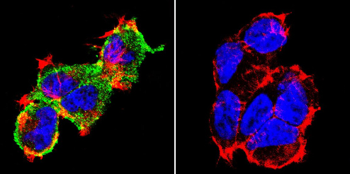 Immunocytochemistry/Immunofluorescence analysis of NFAT2 shows staining in 293 cells. NFAT2 (green), F-Actin staining with Phalloidin (red) and nuclei with DAPI (blue) is shown. Cells were grown on chamber slides and fixed with formaldehyde prior to staining. Cells were incubated without (control) or with ab2796 (1:20) overnight at 4°C, washed with PBS and incubated with a DyLight-488 conjugated secondary antibody. Images were taken at 60X magnification.
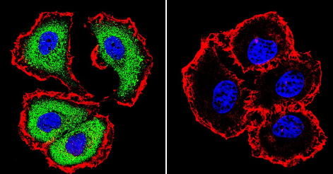
 Immunocytochemistry/Immunofluorescence analysis of NFAT2 shows staining in HeLa cells. NFAT2 (green), F-Actin staining with Phalloidin (red) and nuclei with DAPI (blue) is shown. Cells were grown on chamber slides and fixed with formaldehyde prior to staining. Cells were incubated without (control) or with ab2796 (1:20) overnight at 4°C, washed with PBS and incubated with a DyLight-488 conjugated secondary antibody. Images were taken at 60X magnification.
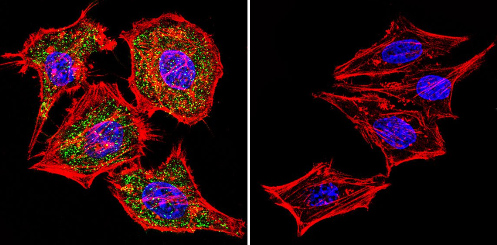
 Immunocytochemistry/Immunofluorescence analysis of NFAT2 shows staining in MCF-7 cells. NFAT2 (green), F-Actin staining with Phalloidin (red) and nuclei with DAPI (blue) is shown. Cells were grown on chamber slides and fixed with formaldehyde prior to staining. Cells were incubated without (control) or with ab2796 (1:20) overnight at 4°C, washed with PBS and incubated with a DyLight-488 conjugated secondary antibody. Images were taken at 60X magnification.
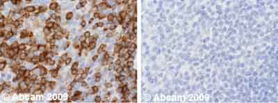 Ab2796 staining Human normal tonsil tissue. Staining is localised to cytoplasm and nucleus.Left panel: with primary antibody at 1 ug/ml. Right panel: isotype control.Sections were stained using an automated system DAKO Autostainer Plus , at room temperature. Sections were rehydrated and antigen retrieved with the Dako 3-in-1 AR buffer EDTA pH 9.0 in a DAKO PT Link. Slides were peroxidase blocked in 3% H2O2 in methanol for 10 minutes. They were then blocked with Dako Protein block for 10 minutes (containing casein 0.25% in PBS), then incubated with primary antibody for 20 minutes, and detected with Dako Envision Flex amplification kit for 30 minutes. Colorimetric detection was completed with diaminobenzidine for 5 minutes. Slides were counterstained with Haematoxylin and coverslipped under DePeX. Please note that for manual staining we recommend to optimize the primary antibody concentration and incubation time (overnight incubation), and amplification may be required.
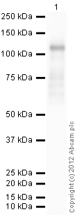 developed using the ECL techniquePerformed under reducing conditions.
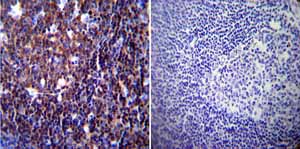 Immunohistochemistry was performed on normal biopsies of deparaffinized Human tonsil tissue. To expose target proteins heat induced antigen retrieval was performed using 10mM sodium citrate (pH6.0) buffer microwaved for 8-15 minutes. Following antigen retrieval tissues were blocked in 3% BSA-PBS for 30 minutes at room temperature. Tissues were then probed at a dilution of 1:200 with a mouse monoclonal antibody recognizing NFATc1 ab2796 or without primary antibody (negative control) overnight at 4°C in a humidified chamber. Tissues were washed extensively with PBST and endogenous peroxidase activity was quenched with a peroxidase suppressor. Detection was performed using a biotin-conjugated secondary antibody and SA-HRP followed by colorimetric detection using DAB. Tissues were counterstained with hematoxylin and prepped for mounting.
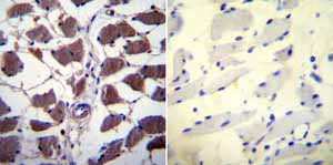 Immunohistochemistry was performed on normal biopsies of deparaffinized Human skeletal muscle tissue. To expose target proteins heat induced antigen retrieval was performed using 10mM sodium citrate (pH6.0) buffer microwaved for 8-15 minutes. Following antigen retrieval tissues were blocked in 3% BSA-PBS for 30 minutes at room temperature. Tissues were then probed at a dilution of 1:20 with a mouse monoclonal antibody recognizing NFATc1 ab2796 or without primary antibody (negative control) overnight at 4°C in a humidified chamber. Tissues were washed extensively with PBST and endogenous peroxidase activity was quenched with a peroxidase suppressor. Detection was performed using a biotin-conjugated secondary antibody and SA-HRP followed by colorimetric detection using DAB. Tissues were counterstained with hematoxylin and prepped for mounting.
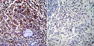 Immunohistochemistry was performed on normal biopsies of deparaffinized Human spleen tissue. To expose target proteins heat induced antigen retrieval was performed using 10mM sodium citrate (pH6.0) buffer microwaved for 8-15 minutes. Following antigen retrieval tissues were blocked in 3% BSA-PBS for 30 minutes at room temperature. Tissues were then probed at a dilution of 1:100 with a mouse monoclonal antibody recognizing NFATc1 ab2796 or without primary antibody (negative control) overnight at 4°C in a humidified chamber. Tissues were washed extensively with PBST and endogenous peroxidase activity was quenched with a peroxidase suppressor. Detection was performed using a biotin-conjugated secondary antibody and SA-HRP followed by colorimetric detection using DAB. Tissues were counterstained with hematoxylin and prepped for mounting.
![Overlay histogram showing Jurkat cells stained with ab2796 (red line). The cells were fixed with 4% paraformaldehyde (10 min) and then permeabilized with 0.1% PBS-Tween for 20 min. The cells were then incubated in 1x PBS / 10% normal goat serum / 0.3M glycine to block non-specific protein-protein interactions followed by the antibody (ab2796, 1µg/1x106 cells) for 30 min at 22ºC. The secondary antibody used was DyLight® 488 goat anti-mouse IgG (H+L) (ab96879) at 1/500 dilution for 30 min at 22ºC. Isotype control antibody (black line) was mouse IgG1 [ICIGG1] (ab91353, 2µg/1x106 cells) used under the same conditions. Acquisition of >5,000 events was performed. This antibody gave a positive signal in Jurkat cells fixed with 80% methanol (5 min)/permeabilized with 0.1% PBS-Tween for 20 min used under the same conditions.](http://www.bioprodhub.com/system/product_images/ab_products/2/sub_3/29857_NFAT2-Primary-antibodies-ab2796-12.jpg) Overlay histogram showing Jurkat cells stained with ab2796 (red line). The cells were fixed with 4% paraformaldehyde (10 min) and then permeabilized with 0.1% PBS-Tween for 20 min. The cells were then incubated in 1x PBS / 10% normal goat serum / 0.3M glycine to block non-specific protein-protein interactions followed by the antibody (ab2796, 1µg/1x106 cells) for 30 min at 22ºC. The secondary antibody used was DyLight® 488 goat anti-mouse IgG (H+L) (ab96879) at 1/500 dilution for 30 min at 22ºC. Isotype control antibody (black line) was mouse IgG1 [ICIGG1] (ab91353, 2µg/1x106 cells) used under the same conditions. Acquisition of >5,000 events was performed. This antibody gave a positive signal in Jurkat cells fixed with 80% methanol (5 min)/permeabilized with 0.1% PBS-Tween for 20 min used under the same conditions.
Overlay histogram showing Jurkat cells stained with ab2796 (red line). The cells were fixed with 4% paraformaldehyde (10 min) and then permeabilized with 0.1% PBS-Tween for 20 min. The cells were then incubated in 1x PBS / 10% normal goat serum / 0.3M glycine to block non-specific protein-protein interactions followed by the antibody (ab2796, 1µg/1x106 cells) for 30 min at 22ºC. The secondary antibody used was DyLight® 488 goat anti-mouse IgG (H+L) (ab96879) at 1/500 dilution for 30 min at 22ºC. Isotype control antibody (black line) was mouse IgG1 [ICIGG1] (ab91353, 2µg/1x106 cells) used under the same conditions. Acquisition of >5,000 events was performed. This antibody gave a positive signal in Jurkat cells fixed with 80% methanol (5 min)/permeabilized with 0.1% PBS-Tween for 20 min used under the same conditions.
Product References
6-Methoxyflavone inhibits NFAT translocation into the nucleus and suppresses T - 6-Methoxyflavone inhibits NFAT translocation into the nucleus and suppresses T
So JS, Kim GC, Song M, Lee CG, Park E, Kim HJ, Kim YS, Jun CD, Im SH. J Immunol. 2014 Sep 15;193(6):2772-83.
Nfatc1 orchestrates aging in hair follicle stem cells. - Nfatc1 orchestrates aging in hair follicle stem cells.
Keyes BE, Segal JP, Heller E, Lien WH, Chang CY, Guo X, Oristian DS, Zheng D, Fuchs E. Proc Natl Acad Sci U S A. 2013 Dec 17;110(51):E4950-9. doi:
Sequential activation of NFAT and c-Myc transcription factors mediates the - Sequential activation of NFAT and c-Myc transcription factors mediates the
Singh G, Singh SK, Konig A, Reutlinger K, Nye MD, Adhikary T, Eilers M, Gress TM, Fernandez-Zapico ME, Ellenrieder V. J Biol Chem. 2010 Aug 27;285(35):27241-50.
Nuclear factor of activated T cells mediates fluid shear stress- and tensile - Nuclear factor of activated T cells mediates fluid shear stress- and tensile
Celil Aydemir AB, Minematsu H, Gardner TR, Kim KO, Ahn JM, Lee FY. Bone. 2010 Jan;46(1):167-75.
Regulation of embryonic kidney branching morphogenesis and glomerular development - Regulation of embryonic kidney branching morphogenesis and glomerular development
Yi T, Tan K, Cho SG, Wang Y, Luo J, Zhang W, Li D, Liu M. J Biol Chem. 2010 Jun 4;285(23):17811-20.
NFAT/Fas signaling mediates the neuronal apoptosis and motor side effects of - NFAT/Fas signaling mediates the neuronal apoptosis and motor side effects of
Gomez-Sintes R, Lucas JJ. J Clin Invest. 2010 Jul;120(7):2432-45.
Overexpression of c-myc in pancreatic cancer caused by ectopic activation of - Overexpression of c-myc in pancreatic cancer caused by ectopic activation of
Buchholz M, Schatz A, Wagner M, Michl P, Linhart T, Adler G, Gress TM, Ellenrieder V. EMBO J. 2006 Aug 9;25(15):3714-24. Epub 2006 Jul 27.
Comparative proteomic studies on the pathogenesis of human ulcerative colitis. - Comparative proteomic studies on the pathogenesis of human ulcerative colitis.
Hsieh SY, Shih TC, Yeh CY, Lin CJ, Chou YY, Lee YS. Proteomics. 2006 Oct;6(19):5322-31.
Expression of NFAT-family proteins in normal human T cells. - Expression of NFAT-family proteins in normal human T cells.
Lyakh L, Ghosh P, Rice NR. Mol Cell Biol. 1997 May;17(5):2475-84.
Two NFAT transcription factor binding sites participate in the regulation of CD95 - Two NFAT transcription factor binding sites participate in the regulation of CD95
Latinis KM, Norian LA, Eliason SL, Koretzky GA. J Biol Chem. 1997 Dec 12;272(50):31427-34.