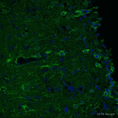
IHC-FoFr image of NG2 staining on sections of rat cortex post injury using ab83178 (1:100). The brain was perfusion fixed using 4% PFA and the sections were permeabilized using 0.1% TritonX in 0.1% PBS. The sections were then blocked using 10% donkey serum for 1hour at 24°C. ab83178 was diluted 1:100 and incubated with the sections for 24 hours at 4°C. The secondary antibody used was donkey polyclonal to rabbit IgG conjugated to Alexa Fluor 488 (1:1000).

Anti-NG2 antibody (ab83178) at 1 µg/ml + Skin (Human) Tissue Lysate - adult normal tissue (ab30166) at 10 µgSecondaryGoat polyclonal to Rabbit IgG - H&L - Pre-Adsorbed (HRP) at 1/3000 dilutiondeveloped using the ECL techniquePerformed under reducing conditions.

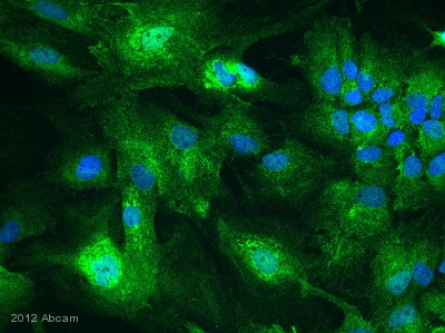
ICC/IF image of ICC/IF staining on miced glia culture cells using ab83178 (1:200). The mixed glia culture was fixed with PFA and the cells were then blocked with 10% donkey serum for 1 hour at 24°C. The cells were then incubated with ab83178 was diluted by 1:200 using 0.3% TritonX with 0.1% PBS and 10% donkey serum. The cells were then incubated with donkey polyclonal to antirabbit conjuaged to Alexa 488 (1:1000). DAPI was used to stain the nucleus.See Abreview


