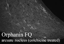
ab10277, at a dilution of 1/500, staining Nociceptin (Orphanin FQ, OFQ) in arcuate nucleus region of rat hypothalamus section by Immunohistochemistry (PFA perfusion fixed frozen sections).

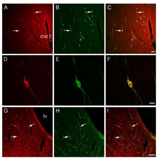
Confocal images showing Nociceptin (OFQ) (red), GnRH (green) and their colocalization (yellow) in the sheep preoptic area and hypothalamus. A-C: Low power view of the preoptic area at the level of the organum vasculosum of the lamina terminalis (OVLT) showing OFQ and GnRH cells, and their colocalization (e.g., white arrows). Bar = 100µm. D-F: High power images of a double-labeled OFQ/GnRH cell; note that the immunofluorescence for OFQ has a punctateappearance in the cytoplasm as opposed to the more diffuse appearance of the GnRH signal. Bar 26 = 20µm. G-I: Single-labeled OFQ cells (e.g., red arrow) in the arcuate nucleus. Note that many GnRH fibers coursing through this region are also OFQ-positive (e.g., white arrows). 3v = third ventricle. Bar = 100µm.

